Download
1 / 12
120 likes | 285 Views
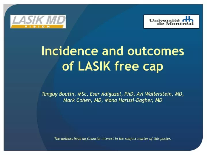
Incidence and outcomes of LASIK free cap . Tanguy Boutin, MSc , Eser Adiguzel, PhD, Avi Wallerstein, MD, Mark Cohen, MD, Mona Harissi-Dagher, MD The authors have no financial interest in the subject matter of this poster. Background.

E N D
Incidence and outcomes of LASIK free cap Tanguy Boutin, MSc, Eser Adiguzel, PhD, Avi Wallerstein, MD, Mark Cohen, MD, Mona Harissi-Dagher, MD The authors have no financial interest in the subject matter of this poster.
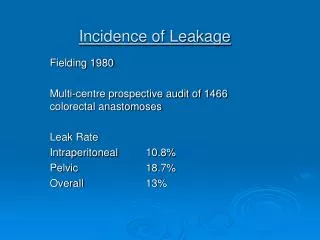
Background Some of the most serious complications of LASIK are linked to the use of the microkeratome.1 The flap can be thin, irregular, buttonhole, incomplete or a dislocated/free cap. Possible causative factors are flat corneas, deep orbits, inadequate suction, decentered ring placement, and faulty microkeratome blades.2
Purpose To determine the incidence, management, and visual outcomes of LASIK free cap following a microkeratome cut.
Methods Retrospective observational case series of 183,941 consecutive LASIK surgeries between 2007 and 2010 in a high volume refractive surgery setting, using standardized equipment and techniques to determine the incidence of intra-operative free cap. Primary outcome measurements: • Manifest Refraction (MR) • Uncorrected distance visual acuity (UDVA) • Corrected distance visual acuity (CDVA) Further corrective surgery was documented as well.
Results 7 eyes of 7 patients were indentified as having an intra-operative free cap out of 183,941 LASIK procedures performed, resulting in an incidence of free cap of 0.004%. Post-operative follow-up: 2 weeks - 27 months. Four of the 7 eyes have long-term follow-up of over 6.5 months. One patient did not have further laser correction, lost 2 lines of UDVA and 1 line of CDVA with mild haze development. Additional corrective surgery was performed in the other three patients, at various times after free cap occurrence. UDVA was greatly increased in those eyes. Only one eye lost one line of CDVA.
Patient 1: 38 year old woman Pre-op 19 months post-free cap
Patient 2: 26 year old man 9 months post-free cap (pre-enhancement) 22 months post-free cap (post-enhancement)
Patient 3: 30 year old woman Pre-op 2 months post-free cap (Pre-PRK)
Patient 4: 31 year old man Pre-op 3.5 months post-free cap (Pre-PRK)
Discussion • The incidence of a free cap complication during LASIK was found to be 0.004% in a 3 year period while other studies have shown an incidence between 0.01% and 1.31%.1,3,4 • Meticulous and precise repositioning of a free cap, using ink reference marks, resulted in minimal loss of CDVA. • The etiology of the causes of LASIK free caps are varied, from flat corneas (K<42D), mispositioning of the microkeratome, suction loss, post-intervention trauma, or unknown etiologies.2,5 • PRK has been used in cases of free cap complications in our group and others resulting in improved UDVA.6
Conclusions • Free cap is a rare intra-operative LASIK flap complication. • This study suggests that immediate and meticulous repositioning of the cap without laser ablation is most appropriate. • PTK/PRK 3 months later is an appropriate refractive surgical management.
References 1. Gimbel et al. Incidence and management of intraoperative and early postoperative complications in 1000 consecutive laser in situ keratomileusis cases. Ophthalmology. 1998;105(10):1839-47. 2. Melki and Azar. LASIK complications: etiology, management, and prevention. SurvOphthalmol. 2001;46(2):95-116. 3. Lin and Maloney. Flap complications associated with lamellar refractive surgery. Am J Ophthalmol. 1999;127(2):129-36. 4. Stulting et al. Complications of laser in situ keratomileusis for the correction of myopia. Ophthalmology. 1999;106(1):13-20. 5. Slade SG. Lasik : Intraoperative (Flap) Complications. In: Alio JL and Azar DT, eds. Management of Complications in Refractive Surgery. Springer, 2008; 15-31. 6. Utz and Krueger. Management of irregular astigmatism following rotationally disoriented free cap after LASIK. J Refract Surg. 2008;24(4):383-91.