Download
1 / 15
210 likes | 508 Views
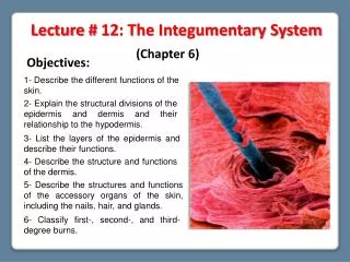
Lecture # 12: The Integumentary System. (Chapter 6) . Objectives:. 1- Describe the different functions of the skin. . 2- Explain the structural divisions of the epidermis and dermis and their relationship to the hypodermis. .

E N D
Lecture # 12: The Integumentary System (Chapter 6) Objectives: 1- Describe the different functions of the skin. 2- Explain the structural divisions of the epidermis and dermis and their relationship to the hypodermis. 3- List the layers of the epidermis and describe their functions. 4- Describe the structure and functions of the dermis. 5- Describe the structures and functions of the accessory organs of the skin, including the nails, hair, and glands. 6- Classify first-, second-, and third-degree burns.
The human body is organized in a hierarchical manner Hierarchical manner: Levels of organization are progressively integrated to make up higher levels. Tissue: It is a combination of similar cells that are grouped together to perform a particular function The human body consists of four basic tissue types: 1- Epithelial tissue 2- Connective tissue 3- Muscle tissue 4- Nervous tissue
T H E I N T E G U M E N T A R Y S Y S T E M 1- The skin and subcutaneous tissue 2- The accessory structures or appendages of the skin Hair Nails Cutaneous exocrine glands
The Skin and Subcutaneous Tissue Functionsof the Skin • 1- Resistance to trauma and infection • 5- Thermoregulation • keratin • acid mantle • thermoreceptors • vasoconstriction / vasodilation • 2- Other barrier functions • 6- Nonverbal communication • waterproofing • UV radiation • harmful chemicals • 3- Vitamin D synthesis • skin first step • liver and kidneys complete process • 4- Sensation • skin is our most extensive sense organ
SKIN Epidermis Dermis Hypodermis Layers of the Skin (from external to internal) 1- Epidermis: is a keratinized, stratified squamous epithelium 2- Dermis: is a dense irregular connective tissue 3- Subcutaneous tissue (Hypodermis): contains adipose tissue
The Epidermis Layers of the Epidermis 5- Stratum corneum It consists up to 30 layers of dead, scaly, keratinized cells, which form a durable surface layer. The surface cells flake off (exfoliate). It is resistant to abrasion, penetration, and water loss 4- Stratum lucidum It is seen only in thick skin thin. It is a translucent zone superficial to stratum granulosum. 3-Stratum granulosum It consists of 3 to 5 layers flat keratinocytes. It contain coarse dark-staining keratohyalin granules 2- Stratum spinosum It consists of several layers of keratinocytes It is the thickest stratum in most skin 1- Stratum germinativum or basale: It is a single layer of cuboidal to low columnar stem cells and keratinocytes resting on the basement membrane The stem cells of stratum basale divide and give rise to keratinocytes that migrate toward skin surface and replace lost epidermal cells
The Epidermis Cells of the Epidermis • They are macrophages originating in bone marrow that guard against pathogens. They are found in stratum spinosum and granulosum where they stand guard against toxins, microbes, and other pathogens that penetrate skin 5- Dendritic (Langerhans) cells 2- Keratinocytes: • The great majority of epidermal cells. They synthesize the keratin Exfoliating keratinocytes Dead keratinocytes Living keratinocytes 4- Tactile (Merkel) cells • They occur in the basal layer of epidermis. They are touch receptor cells associated with dermal nerve fibers 3- Melanocytes • They occur only in stratum basale. They synthesize the pigment melanin that shields DNA from ultraviolet radiation 1- Stem cells • Undifferentiated cells that give rise to keratinocytes. They are in the deepest layer of epidermis (stratum basale)
The Life History of a Keratinocytes In 30 - 40 days a keratinocyte makes its way to the skin surface and flakes off Newly formed keratinocytes push the older ones toward the surface Keratinocytes are produced deep in the epidermis by stem cells in stratum basale • This migration is slower in old age, and faster in skin injured or stressed
The Dermis Epidermal ridges Layers of the Dermis 1- Papillary layer Dermal papillae It is the superficial zone of dermis. It consists of a thin zone of areolar tissue in and near the dermal papilla. It allows for mobility of leukocytes and other defense cells should epidermis become broken. It is rich in small blood vessels They are upward fingerlike extensions of the dermis. The dermal papillae and the epidermal ridges determine the fingerprints It supports and nourishes the overlaying epidermis 2- Reticular layer It is deeper and much thicker layer of dermis. It consists of dense irregular connective tissue, which contains arteries, veins, sweat and sebaceous glands, and pressure receptors (Pacinian corpuscles). The reticular layer helps resist tension in the skin.
Epidermis Dermis Hypodermis The Hypodermis • It is the subcutaneous tissue. It has more areolar and adipose than the dermis Functions: • 1- It pads the body • 2- It binds skin to underlying tissues • 3- Drugs are introduced by injection in the hypodermis, which is • highly vascular & absorbs them quickly • 4- It contains the subcutaneous fat, which is an energy reservoir, and acts as thermal insulation
Hair The portion above the skin surface Hair shaft Follicle It is a diagonal tube that dips deeply into dermis and may extend into hypodermis • It is a bundle of smooth muscle cells. In response to cold, fear or other stimuli, contracts and make the hair stand on end Piloerector muscle The remainder of the hair in the follicle Hair root • It is the region of mitotically active cells immediately above papilla. It is the hair’s growth center Hair matrix • It is swelling at the base where hair originates in dermis or hypodermis. Only living hair cells are in or near bulb Hair bulb • It is a bud of vascular connective tissue encased by bulb. It provides the hair with its sole source of nutrition Blood capillaries in dermal papilla
Functions of Hair: • Most hair on trunk and limbs is vestigial • little present function • warmth in ancestors • 1- Hair receptors alert us of parasites crawling on skin • 2- Scalp helps retain heat • 3- Scalp protects against sunburn • 4- Gender identification • 5- Pubic and axillary hair signify sexual maturity and aids in transmission of sexual scents • 6- Guard hairs (vibrissae) guard nostrils and ear canals. The eyelashes shield the eyes • 7- Eyebrows are important in nonverbal communication Cuticle Cortex Medulla
Nails Free edge They are modification of the epidermis, which cover and protect the dorsal surface of the distal part of fingers. They contain hard keratin. Nail body Nail plate Nail groove Nail fold Eponychium (cuticle) Lunule It is the skin underlying the nail plate Nail bed It is the growth zone of thicken stratum basale at the proximal end of nail. Mitosis here accounts for nail growth Nail matrix
Cutaneous Glands • 1- Merocrine (eccrine) sweat glands 2- Apocrine sweat glands • They are the most numerous skin glands - 3 to 4 million in adult skin. They are simple tubular glands, which ducts open by way of a pore on the skin surface. • They occur in groin, anal region, axilla, areola, bearded area in mature males. • They have ducts that lead to nearby hair follicles. They produce a watery perspira- tion that helps cool the body • They develop at puberty and produce a sweat that is thicker, milky, and contains fatty acids. They are scent glands that respond to stress and sexual stimulation. 3- Sebaceous glands They are flask-shaped glands with short ducts opening into hair follicle, which produce the sebum (an oily secretion) • They are holocrine gland: their secretion consists of broken down cells, which are replaced by mitosis at base of gland • The sebum keeps skin and hair from becoming dry, brittle, and cracked Holocrine gland
Classification of Burns Partial-thickness burns Full-thickness burns (a) First degree (b) Second degree (c) Third degree • They involve only the epidermis. • They are marked by redness, slight edema, and pain. They heal in a few days. • Ex: Most sunburns are first degree burns • They involve the epidermis and part of the dermis. • They produce blisters and are very painful. They leave at least part of the dermis intact. They may be red, tan, or white. • They heal from two weeks to several months may leave scars. They involve the epidermis and all of the dermis, and often some deeper tissues (muscles or bones) are destroyed • They often require skin grafts, fluid replacement and infection control


