Download
1 / 39
450 likes | 1.72k Views
UNIT –II COMMON SIGNS & SYMPTOMS TOPIC: FLUID IMBALANCES. Prepared by, Mrs. Anju Ullas Lecturer Dept. of Medical Surgical Nursing Yenepoya Nursing College. Learning objectives Students will be able to: explain the regulation of water balance

E N D
UNIT –II COMMON SIGNS & SYMPTOMS TOPIC: FLUID IMBALANCES Prepared by, Mrs. AnjuUllas Lecturer Dept. of Medical Surgical Nursing Yenepoya Nursing College
Learning objectives Students will be able to: • explain the regulation of water balance • define the meaning of extracellular and intracellular fluid compartments • enlist and explain the different types of fluid imbalances
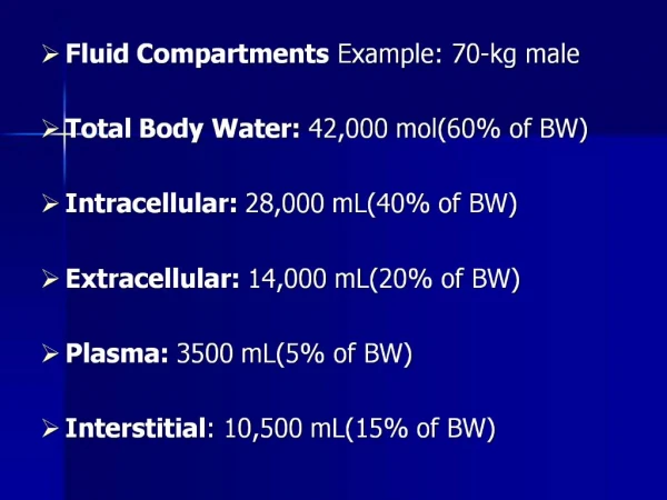
Introduction: • Every part of your body needs water to function. When you are healthy, your body is able to balance the amount of water that enters or leaves your body. • Water is the major body component, accounting 60% of the adult body weight. • 2/3rd of the water is with in the cells. (intracellular fluid) • 1/3rd of body water is outside the cells(extracellular fluid).
The extra cellular fluid compartment is further divided into • intravascular (eg:plasma) • interstitial(between cells).
Regulation of body fluid volume Hypervolemia Hypovolemia inhibits stimulates thirst, release of ADH& Aldosterone thirst, release of ADH&Aldosterone Increased urination of Decreased urination dilute urine of concentrated urine Normal fluid volume restored
Types of fluid imbalances 5 major types • Extracellular fluid volume deficit • Intra cellular fluid volume deficit • Extra cellular fluid volume excess • Intra cellular fluid volume excess • Extracellular fluid volume shift.
1.Extra cellular fluid volume deficit(dehydration): It is a decrease in intravascular and interstitial fluids. • Etiology :- • Lack of fluid intake • Excess fluid losses -severe vomiting and diarrhea. • other potential causes are fever, burns, blood loss, Increased ADH secretion, use of diuretics, diuretic phase of acute renal failure
Pathophysiology:- causes and risk factors fluid loss from intravascular spaces compensatory mechanism ADH and Aldosterone secretion increased to reabsorb water and sodium in the kidney. Fluids are also absorbed from the ileum and large colon.
Baroreceptors sense low BP. Stimulation of sympathetic nervous system increase heart rate movement of fluid from periphery to circulation if compensatory mechanism fails. Dehydration
Types:- • Hyperosmolar fluid volume deficit water loss greater than electrolyte loss. • Iso-osmolar fluid volume deficit both losses are equal. • Hypotonic fluid volume deficit electrolyte loss greater than fluid loss
Clinical manifestations • Loss of body weight • Changes in intake and output. • Increased thirst • Decreased pulse • Manifestations of cellular dehydrationdry mouth and eyes, decreased skin turgor, soft and sunken eyes, muscle weakness, constipation . • cerebral signs:-restlessness, head ache, confusion, followed by coma.
Management:- Fluid restoration • Oral rehydration • Intravenous rehydration • Correction of underlying problem with antiemetics, antidiarrheals, antibiotics, and antipyretics.
Nursing management:- Assessment:- • Obtain the client history of fluid loss • Check the vital signs every 2-4 hrs. • Assess orthostatic BP and pulse changes
Monitor intake –out put and daily weight • Monitor plasma sodium ,BUN, glucose,andhematocrit levels. • Assess the oral cavity for dryness of mucus membrane. • Check skin turgor • Generalized weakness may develop because changes in Na levels.
Nursing diagnosis:- • Fluid volume deficit related to vomiting, diarrhea, hemorrhage, or third space fluid loss such as ascites or burns. • Altered mucus membranes related to lack of oral intake, or other causes. • Risk for injury related to orthostatic hypotension.
2.Intra cellular fluid volume deficit:- • It occurs quite often in older clients • Thirst and oliguria are the most common compensatory signs. • Cellular manifestations fever, CNS changes such as confusion, coma, and cerebral hemorrhage. • Rx:- I/V fluids, correction of underlying cause.
3. Extracellular fluid volume excess:- • ECF volume excess is fluid overload or overhydration. • It can be seen in Vascular system hypervolemia Interstitial space third spacing Etiology:- it can develop from two processes. a. Simple overloading with fluids b. Failure to excrete fluids.
Pressures • hydrostatic pressure pushing fluid and solute OUT of the capillaries, • oncotic pressure (also known as colloid osmotic pressure) pulls fluid into the capillaries and prevents fluid from leaving.
Pathophysiology:- • Fluid overload Increased hydrostatic pressure in arterial end of capillary Increased peripheral Vascular resistance Fluid movement into tissue Increased left atrial & Ventricular pressure Edema LVF, PE
B. Decreased serum albumin Decreased production of plasma protein Decreased capillary oncotic pressure Edema
c. Tissue injury Increased capillary permeability Movement of plasma protein into tissues Increased tissue oncotic pressure Edema
Clinical manifestation:- Respiratory manifestations: • Coughing • Dyspnoea • crackles over affected area • Pallor, cyanosis • decreased tissue perfusion • If hydrostatic pressure continues to rise fluid shifts into the pleural space leads to pleural effusion.
Cardiac manifestations: • distended jugular vein • a bounding pulse and elevated blood pressure • increased CVP • heart sound S3 can often be auscultated.
Edema of the feet • rapid weight gain(a classical sign of fluid overload) • CNS changes include confusion and head ache. • As the fluid excess increases lethargy occures, followed by seizures and coma.
Management:- • Restriction of sodium and fluids. • Promoting urine output: Mild diuretics and digitalis promote urine output and myocardial contractility. • Nursing management:- • Assessment: • Monitor vital signs for bounding pulse, elevated BP. • Assess breath sounds every 4-8 hrs for crackles, wheezes, rhonchi.
Compare I/O every 4-8 hrs • Weigh the client daily. • Monitor, sodium level, hematocrit, and urine special gravity. • Observe changes in LOC. • Nursing diagnosis:- • Fluid volume excess related to heart ,renal or liver failure.
4. Intra cellular fluid volume excess Etiology:- • water excess or solute deficit • Most common cause during hospitalization ; - administration of hypo osmolar I/V fluids such as 0.45%NS ,5% dextrose in water. • people with certain psychiatric disorders ,such as schizophrenia
Pathophysiology: Movement of fluid from lower concentration to high concentration by osmosis to maintain equilibrium. cellular edema • Cerebral cells absorb the fluids more quickly than other cells. Cerebral edema
Clinical manifestations • Weight gain • Non pitting edema • Seizures • Coma • Unconsciousness
Management: • Reduce ICP with osmotic diuretics. • Perform neurologic assessment. • Monitor I/V fluids and I/O hourly ,daily weight. • Provide safety measures to protect the patient.
5. Extracellular fluid volume shift : • Fluid shift are of two types: • Vascular to interstitial spaces leads to fluid volume deficit (hypovolemia) • Interstitial to vascular space leads to fluid volume excess (hypervolemia). Common sites of third spacing: pleural cavity, peritoneal cavity, and pericardial sac.
Etiology:- • Increased capillary permeability, • increased hydrostatic pressure • Any tissue injury:- eg:- crush injuries, major surgery, burns, bowel obstruction, and sepsis
Management:- Based on the cause. For eg:- fluid collects b/o pericarditis- pericardiocentasis bowel obstruction ->paracentesisthoracentesis • Replace fluids : a large volume of Iso-osmolar I/V fluid administration is required to replace intravascular volume.
Conclusion Fluid balance can alter with disease and illness so it important to be aware of how much fluid is in the body, taking steps such as measuring urea and electrolytes levels and how to balance fluid in the body
Summary In this topic we have discussed about the regulatory mechanism of fluids in the body and the types of fluid imbalances, which include definition, causes, pathophysiology, clinical manifestation and how to overcome the imbalances (management).
Evaluation • What is mean by fluid imbalance? • How does the regulation of water balance takes place in body • Explain extracellular and intracellular fluid compartments • Enlist and explain the different types of fluid imbalances
References:- • Black MJ.Textbook of medical surgical nursing.7thed.St. louis:Saunders • Brunner.Text book of medical surgical nursing.6thed.Philadelphia:Saunders; • Lewis.Medical surgical nursing.6th ed. St louis:Mosby • http://www.journal of infusion nursing.com/pt/re/currentoc.htm. • http://www.journal of parenteral and enteral nutrition.com/p/articles/mi