Download
1 / 72
720 likes | 961 Views
Hypothyroidism. Iodine deficiency disorders. Department of Internal Medicine N2 as.-prof. Svystun I. I. Anatomy of thyroid gland. The thyroid is a firm vascular organ lying in the neck, caudal to cricoid cartilage.

E N D
Hypothyroidism. Iodine deficiency disorders. Department of Internal Medicine N2 as.-prof. Svystun I. I.
Anatomy of thyroid gland The thyroid is a firm vascular organ lying in the neck, caudal to cricoid cartilage. It is composed of two nearly equal lobes connected by a thin isthmus and weights approximately 20 – 30 g. Rests of thyroid tissue are occasionally presents in sublingual or retrosternal areas. Thyroid secrets: T3, T4, calcitonin. Hyoid Bone Cricothyroid Ligament Thyroid Cartilage Cricoid Cartilage Thyroid Gland Pyramidal Lobe Right Lobe Isthmus Left Lobe Trachea
A follicle is structural and functional unit of the thyroid gland • The follicle contains colloid (which consists of thyroglobuline, a glycoprotein, containing T3 and T4 within its matrix) • Epithelial cells • Parafollicle cells (C-cells) (synthesized calcitonine) people.upei.ca
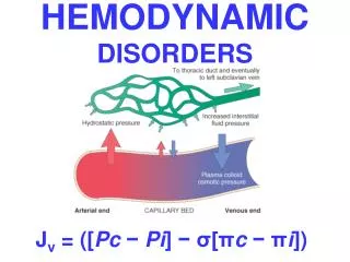
The thyroid hormones, thyroxine (T4) and triiodothyronine (T3) are secreted under the stimulatory influence of pituitary thyrotropin (thyroid-stimulating hormone or TSH). TSH secretion is primary regulated by a dual mechanism: • thyrotropin-releasing hormone (TRH); • thyroid hormone. Thyroid hormone exits in circulation in both free and bound formas. The thyroid gland is the sole source of T4 and only 20% of T3 is secreted in the thyroid. Approximately 80% of T3 in blood is derived from peripheral tissue (mainly hepatic or renal) deiodinatoin of T4 to T3. Hypothalamus TRH stimulation inhibition Pituitary Free hormone Bound hormone T4 T3 Liver Thyroid Binding proteins TSH stimulation Thyroid
Thyroid hormone synthesis howmed.net
Major deiodinative and non-deiodinative pathways of thyroid hormones metabolism. http://health-7.com/
Physiologic effects of thyroid hormones • Increasing of protein metabolism in virtually every body tissue • Increasing of O2 consumption by increasing the activity of Na+ H+ ATPase (Na pump), primarily in tissues responsible for basal O2 consumption (i.e., liver, kidney, heart and skeletal muscle).) • Stimulation of erythropoesis • Positive chrono- inotropic effects on myocardium • Achievement of formation of nervous system and skeleton in perinatal period
Goitre • is an enlargement of the thyroid gland.
Hypothyroidism (myxedema) is the characteristic reaction to thyroid hormone deficiency. The spectrum of hormone ranges from a few non – specific symptoms to overt hormone, to myxedema coma.
Historical perspective • The first full clinical description of the hypothyroidism and mixedema was made in 1874 by Gull (by the cretinoid state supervening in adult life in women) • Term “myxedeme” first used by Ord in 1978
Epidemiology -Hypothyroidism occurs in 3 to 6 % for the adult population, but is symptomatic only in a minor of them. • Usually develops after the age of 30 • It occurs 8 to 10 times more often in women than in men
I. Congenital II. Acquired 1. Laboratory (subclinical) hypothyroidism 2. Clinical hypothyroidism 1. Primary (thyroid gland disturbances). 2. Secondary (due to pituitary disease). 3.Tertiary (due to hypothalamic disease). 4.Peripheral. Classification
Etiology of primary hypothyroidism • Congenital • Maldevelopment –hypoplasia or aplasia • Inborn deficiencies of biosynthesis or action of thyroid hormone • Atypical localization of thyroid gland • Severe iodine deficiency • Acquired • iodine deficiency • autoimmune processes (Hashimoto’s thyroiditis): MAE 1 & 2 • surgical subtotal removal or total thyroidectomy • irradiation therapy (organs of the neck) • I131 therapy • during or after therapy with propylthyouracil, methimazole, iodides for hyperthyroidism • infiltrative diseases (tuberculosis, actynomycosis) • trauma • medications such as amiodarone, interferon alpha, thalidomide
Etiology of secondary and tertiary hypothyroidism • Congenital - Congenital pituitary or hypothalamus disorders (anatomical or physiological) • Acquired • Tumors or metastasis • hemorrhagic necrosis (Sheehan’s syndrome) • Inflammatory disorders (infection, infiltrative process) • Trauma • surgical and radiation treatment for pituitary or hypothalamic disease • Chemical and pharmacological intoxications (reserpin, bromocriptine).
Etiology of peripheral hypothyroidism • peripheral tissue resistance to thyroid hormones • decreasing of T4 peripheral transformation into T3 (in liver or in kidneys) • production of antibodies to thyroid hormones
Skin and hair • Skin is dry, thick and silk, is often cool and pale. • Nonpitting edema of the hands, feet and periorbital regions (myxedema). Pitting edema also may be present. • The faces are puffy and features are coarse. the loss of the lateral aspect of the eyebrow, sometimes termed Queen Anne's sign
Skin and hair • Skin may be orange due to accumulation of carotene. • Hair may become course and brittle, hair growth slows and hair loss may occur. Lateral eyebrows thin out and body hair is scanty. • Hypothyroidism does not cause obesity, but modest weight gain from fluid retention and fat deposition often occurs
Nervous system Patients complain on fatigue, loss of energy, lethargy, forgetfulness, reduced memory. • Their level of physical activity decreases, and they may speak and move slowly. Mental activity declines and there is inattentiveness, decreased intellectual function, and sometimes may be depression. • Neurological symptoms include also hearing loss, parasthesias, objective neuropathy, particularly the carpal tunnel syndrome, ataxia. • Tendon reflex shows slowed or hung-up relaxation.
Cardiovascular system Complains on: dyspnea, pain in the region of the heart Objective examination: • Increased peripheral resistance • Hypertension • Bradycardia • LV hypertrophy with decreased contractility, reduced cardiac output • Pericardial effusion • Congestive heart failure • The ECG may show low voltage and/or non-specific ST segment and T wave changes. • Hypercholesterolemia
Gastrointestinal system • Gastrointestinal motility is decreased loading to constipation and abdominal distension, pseudoobstruction of intestines, paralytic ileus. • Abdominal distension may be caused by ascities as well. Ascitic fluid, like other serous effusions in myxedema, has high protein content. • Achlorhydria occurs, often associated with pernicious anemia.
Renal system • Reduced excretion of a water load may be associated with hyponatriemia Renal blood flow and glomerular filtration rate are reduced, but serum creatinine is normal May be mild proteinuria and infections of urinary tract Respiratory system • Dyspnea of effort is common. • This complaint may be caused by enlargement of the tongue and larynx, causing upper airway obstruction, or by respiratory muscle weakness, interstitial edema of the lungs, and for plural effusions which have high protein content • Hoarseness from vocal curt enlargement often occurs
Musculoskeletal system • Muscle and joint aches, pains and stiffness are common • Objective myopathy and joint swelling or effusions are less often present • The relaxation phase of the tendon reflexes is prolonged • Serum creatine phosphokinase and alanine aminotransferase activities are often increased, probably as much to slowed enzyme degradation as to increased release from muscle
Blood disorders • Anemia, usually normocytic, caused by decreased red blood cell production, may occur. It is probably from decreased need of peripheral oxygen delivery rather than hematopoetic defect • Megaloblastic anemia suggests coexistent pernicious anemia • Most patients have no evidence iron, folic acid or cyancobalamin deficiency
Endocrine system • Thyroid gland: nonpalpable or enlargement. • Adrenal glands: hypofunction • Pituitary system: secretion of growth hormone is deficient because thyroid hormone is necessary for synthesis of growth hormone. Growth and development of children are retarded. Epiphyses remain open. • Gonadal glands: menorrhagia (from anovulatory cycles), secondary amenorrhea, infertility and galactorrhea; decreased potention in men
Metabolic system • Hypothermia is common • Hyperlipidemia with increase of serum cholesterol and trigliceride occurs because of reduced lipoprotein lipase activity
Clinical features • Hypothyroidism can be presented in many different ways and can mimic other disorders • Because many manifestations of hypothyroidism are non-specific, the diagnosis is particularlylikely to be overlooked in patients with otherchronic illnesses and elderly and can lead to significant morbidity and even mortality
Subclinical (laboratory) hypothyroidism It is a state in which we can’t find clinical features of hypothyroidism and euthyroidism is reached by compensatory increasing of TSH secretion and that’s why synthesis and secretion of such level of thyroid hormone that will be enough for organism. It is an asymptomatic state in which serum T4 and free T4 are normal, but serum TSH is elevated. This designation is only applicable when thyroid function has been stable for weeks or more, the hypothalamic-pituitary-thyroid axis is normal, and there is no recent or ongoing severe illness.
Diagnosing and Managing Thyroid Disease in the Nursing Home . JAMDA. Volume 9, Issue 1 , Pages 9-17, January 2008
Recommendations of Six Organizations Regarding Screening of Asymptomatic Adults for Thyroid Dysfunction
Replacement therapy • Synthetic preparations • T4 (l-thyroxine) • T3 (liothyronine sodium) • Combined: • Thyrocomb • Thyrotom • Thyroidin • Novothyral Side effects • Allergic reactions • Angina pectoris • Cardiac arrhythmia
Congenital hypothyroidism • Children are born with increased weight • Subcutaneous edema • Hypotermia • Prolonged jaundice • Physical (dwarfism) and mental retardation (cretinism)
Peculiarities of treatment < 3 month – 25 mcg/day 3 -12 month – 37,5 mcg/day 1 – 5 years – 75 mcg/day 5 – 7 years – 75 – 100 mcg/day > 7 years – 100 mcg/day
Myxedema coma - is a life-threatening complication of hypothyroidism Precipitating factors include • exposure to cold • infection • Trauma • Surgery • Myocardial infarction • Bleeding • Stress situation • Drugs that suppress the CNS
Clinical signs of myxedema coma • Slow development (weakness, somnolence, coma) • extreme hypothermia (temperatures 24 to 32) • Areflexia • Seizures • Bradycardia, hypotension • Polyserositis • CO2 retention, and respiratory depression caused by decreased cerebral blood flow, nonreversible brain changes • Rapid diagnosis (based on clinical judgment, history, and physical examination) is imperative because early death is likely.
Treatment of myxedema coma • large doses of T4 (200-500 mcg i/v bolus 3 – 4 times a day) or T3 if available (40 – 100 mcg i/v bolus 3 times a day), because TBG must be saturated before any free hormone is available for response. • The maintenance dose for T4 is 50 mkg/day i/v and for T3 10 -20 mcg/day i/v until the hormone can be given orally.
Treatment of myxedema coma • Corticosteroid therapy (hydrocortisone 200 – 400 – 600 mg/day i/v). • The patient should not be rewarmed rapidly because of the threat of cardiac arrhythmia. • Hypoxemia is common, so PaO2 should be measured at the outset of treatment. If alveolar ventilation is compromised, immediate mechanical ventilatory assistance is required.
Thyroiditis The various types of thyroiditis encompass a heterogeneous group of inflammatory disorders of diverse etiologies and clinical features. With all forms of thyroiditis, destruction of the normal architecture of the thyroid follicular occurs, yet each disorder has distinctive histologic characteristics.
Classification • Acute thyroiditis. • Subacute thyroiditis: • subacute granulamatous thyroiditis; • subacute lymphocytous thyroiditis. • Chronic thyroiditis: • Hashimoto thyroiditis; • Ridel struma. • Specific thyroiditis. • Thyroiditis caused by mechanical or physical factors.
Etiology: a bacterial pathogen: St. aureus, Str. hemolyticus, Str. pneumonie, anaerobic organisms, E. coli, coccidiodomycosis. Infection occurs either secondary to hematogenous or lymphatic spread, or direct introduction of an infective agent by trauma. Clinical signs: fever, chills and other signs of abscess formation. Anterior neck swelling and pain radiating to the ear or mandible. The physical examination suggests the presence of an abscess, with erythema of the skin, marked tenderness to palpation, and at times fluctuance. Laboratory: Leucocytosis with a left shift, increased ESR. Thyroid hormone concentrations in blood are normal, although hyperthyroxinemia has been reported Acute thyroiditisis an acute bacterial inflammation
Treatment • Patient should be treated at surgical department. • Parental antibiotics should be administered according to the specific pathogen identified. • If fluctuance is present, incision and drainage might be required. • Bacterial thyroiditis must be treated early and aggressively, since abscess formation can occasionally dissect downward into the mediastinum. Recurrences of the disorder are very rare. • (Duration of the treatment must be nearly 1,5-2 month).
Subacute thyroiditis –an acute inflammatory disease of the thyroid probably caused by a virus with destruction of thyrocytes • subacute granulamatous thyroiditis; • subacute lymphocytous thyroiditis
Etiology Coxsackie virus Adenovirus Mumps Echovirus Influenza Epstein-Barr viruses A genetic predisposition is likely because of the association of HLA-BW 35 histocompatibility antigens. Clinical signs unilateral anterior neck pain, often associated with unilateral radiation of pain to the ear or mandible. Pain is often proceeded by a few weeks prodrome of myalgias, low-grade fever, malaise and sore throat. Dysphagia Symptoms of hyperthyroidism Physical examination:an exquisitely tender, very hard, nodular enlargement, which is most often unilateral. Tenderness is often so extreme that palpation is limited. Tachycardia, a widened pulse pressure, warm skin and diaphoresis are also observed when hyperthyroidism is present. Subacute thyroiditis
Subacute thyroiditis • Laboratory findings • Early in the disease we can find an increase in T4, a decrease in RAI uptake (often 0), leucocytosis and a high ESR. • After a several weeks, the T4, is decreased and the RAI uptake remains low. • Full recovery is the rule; rarely, patients may become hypothyroid. • Treatment - An acute phase lasts from 4-8 weeks, during which treatment is symptomatic (aspirin 600 mg q 3-4 h, prednisolone 10-20 mg orally tid; after 1 week prednisolone can be tapered by 5 mg every 2-3 days; thus glucocorticoids are usually not required for longer than several weeks. • Symptomatic therapy due to the phase (hyperthyroid – anrithyroid drugs, beta-blockers, hypothyroid – thyroid hormone replacement (levothyroxine 100-150 mkg/day). • Following the hypothyroid phase recovery occurs, and the normal histologic features and secretory capacity of the thyroid are restored.
Ultrasound of the right lobe of the thyroid demonstrates an ill-defined irregular region of heterogeneous hypoechogenicity without elevation of flow on Colour Doppler examination. http://radiopaedia.org