Download
1 / 79
810 likes | 858 Views
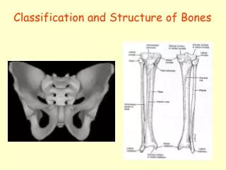
CLASSIFICATION OF BONES. BONE REVIEW. A bone is a specialized type of connective tissue. Bone is distinguished from other CT by the mineralization of its matrix, leading to the production of hard tissue. There are 206 bones in the body Bone tissue is classified

E N D
BONE REVIEW • A bone is a specialized type of connective tissue. • Bone is distinguished from other CT by the mineralization of its matrix, leading to the production of hard tissue. • There are 206 bones in the body • Bone tissue is classified • According to development: membranous and cartilaginous bones • According to shape: long, short, flat, irregular and sesamoidbones • According to region of location in the body: Axial and appendicular bones
ACCORDING TO DEVELOPMENT • Membranous bones: they develop through intramembranous ossification • Eg: clavicle, bones of the face and vault of the skull • Cartilaginous bones: they develop through endochondral ossification. • Here a pre existing cartilaginous model of a bone is gradually destroyed and replaced by a bone • Eg: long bones of the upper and lower limbs
ACCORDING TO SHAPE (e) Sesamoid bone: patellae
ACCORDING TO SHAPE • Classified based on the shape. The location of the spongy and compact tissues of the bone varies with the shape of the bone LONG BONES • Long bones: are longer than they are wider. • Consist of a shaft and two ends. • Thick compact tissue form the outer coat • While spongy tissue is seen internal to the compact bone and at the two ends • Eg bones of the upper and lower arms and legs as well as the metacarpals and metatarsals.
ACCORDING TO SHAPE SHORT BONES • Short bones: Roughly cube (cuboidal) shaped. • They have only a thin Layer of compact bone tissue surrounding a spongy interior eg carpal and tarsal bones FLAT BONES • Are thin, flattened and plate like. • Consist of two layers of thick compact bones with an intervening layer of spongy bone • Eg: sternum, scapulae, cranium.
ACCORDING TO SHAPE IRREGULAR BONE • Are irregular and complex in shape. • Consist of thin layer of compact bone surrounding a spongy interior • The bone may contain air spaces (pnumatic bones) or sinuses (ethmoid bone) • eg vertebrae, hip bone SESAMOID BONE (bone in the tendon) • They are bones embedded in tendons. They are associated with tendons • Eg: Patellae (patella tendon), pisiform bone (tendon of FCU).
ACCORDING TO REGION • AXIAL BONES: they belong to the axial skeleton and are found in the region of axial skeleton • Eg: bones of the skull, vertebrae, auditory ossicles, sternum and ribs • APPENDICULAR BONES: they belong to the appendicular skeleton and are found in the region of the appendicularskeletion • Eg: bones of the limbs and girdles (pelvic and pectorial girdles)
BONE MARKINGS AND FEATURES • Bone markings are marks and features that appear where: • Tendons, ligaments, fascia are attached to the bone • Arteries lie adjacent to or enter the bone • Some of the bone markings and features are: • -Capitulum: small round articular head. Eg: capitullum of humerus. • -Condyle: rounded knuckle-like articular area, usually occurring in pairs. Eg: lateral femoral condyle. • -Crest: ridge of bone eg: the iliac crest of hip bone. • Epicondyle: eminence superior to a condyleeg: lateral epicondyle of humerus
BONE MARKINGS AND FEATURES • Facet: smooth flat area, usually covered with cartilage where a bone articulate with another bone. Eg: superior castal facet on the body of a vertebra for articulation with a rib. • Foramen: passage through a bone eg: obturator foramen. • Fossa: hollow or depressed area. Eg: infraspinousfossa of the scapular. • Groove: elongated depression or furrow, eg radial groove of the humerus. • Head: large round articular end. Eg: head of humerus • Line: linear elevation eg: soleal line of the tibia
BONE MARKINGS AND FEATURES • Malleolus: Rounded process. Eg: lateral malleolus of fibula. • Notch: indentation at the edge of a bone. Eg: greater sciatic notch. • Protuberance: projection of a bone. Eg: external occipital protuberance. • Spine: thorn-like process. Eg: the spine of the scapular. • Spinous process: projecting spine-like part. Eg: spinous process of a vertebra.
BONE MARKINGS AND FEATURES • Trochlea: spool-like articular process that acts as a pulley. Eg: trochlea of the humerus. • Trochanter: larhe blunt elevation. Eg: the greater trochanter of femur. • Tubercle: small raised eminence. Eg: greater tubercle of the humerus. • Tuberosity: large rounded elevation. Eg: ischialtuberosity.
THE ARM • The arm extends from the shoulder joint to the elbow joint. • The humerus is the bone of the arm It contains • four muscles, • Nerves (musculocutaneous, readial, ulnar and median nerves) and • Blood vessels.
COMPARTMENTS OF THE ARM • The medial and lateral intermuscular septa (extension of deep fascia) divide the arm into: • Anterior compartment • Posterior compartments • The intermuscular septa are attached to the medial and lateral borders and supracondylar ridges of the humerus
COMPARTMENTS OF THE ARM • The septa functions to • provide extra surface for attachments of muscles • Provide plane alone which nerves and blood vessels run • Medial septa is pierced by the ulnar nerve and sup. ulnar collateral artery • While the lateral septum is pierced by the radial nerve and anterior descending branch of profundabrachii artery
ANTERIOR COMPARTMENT OF ARM Three flexor muscles out of the four major arm muscles are in the anterior (flexor) compartment. These muscles are: Coracobrachialis Biceps brachii Brachialis
ANTERIOR COMPARTMENT OF ARM cont’d Gray’s Anatomy, 4th edition, (Susan, 2008)
CORACOBRACHIALIS • Origin: Apex of the coracoids process. • Insertion: Midway along the medial border of the humerus • Nerve supply: Musculocutaneous nerve (C5,6). • Action: Weak flexor and adductor of the shoulder joint. Diag: clin Anat,6thed (moore, 2010)
CORACOBRACHIALIS • Coracobrachialis is an elongated muscle in the superomedial part of the arm. • It is an important landmark for locating other structures in the arm. • Coracobrachialis helps flex and adduct the arm and stabilize the glenohumeral joint
BICEPS • Biceps femoris has two heads • Origin: Long head from the supraglenoid tubercle and adjoining part of the glenoid labrum of the scapula. Short head from the apex of the coracoid process (with the coracobrachialis). • Insertion: radial tuberosity via the biceps tendon. The tendon has a broad medial expansion called bicipitalaponeurosis, which merges with the deep fascia of the arm and with the capsule of the elbow joint • Nerve supply:Musculocutaneous nerve (C5,6) with one branch to each belly. • Action: Powerful flexor of the elbow and supinator of the forearm. Diag: clinAnat, 6thed, (Moore,2010)
BICEPS cont’d • Biceps is a ‘’three-joint muscle’’ that has no attachment to the humerus. • The action and effectiveness of biceps are markedly affected by the position of the elbow and the forearm. • The rounded tendon is surrounded by a sheath of synovial membrane as it descends in the intertuberculersulcus of the humerus Diag: clinAnat, 6thed, (Moore,2010)
BICEPS cont’d • The two bellies lie side by side and are connected loosely by areolar tissue, but do not merge until just above the elbow joint . • The flattened tendon at the lower (distal) part or end rotates with anterior surface turning laterally as it passes through the cubitalfossa to its insertion • The tendon has a broad medial expansion called the bicipitalaponeurosis Diag: clinAnat, 6thed, (Moore,2010)
BICEPS cont’d Diag: clin Anatomy by region,9thed, (Snell,2012)
BICEPS BRACHI TEST • Test: The forearm is supinated and the elbow is flexed against resistance. The contracted muscle will form a prominent bulge, and the tendon and aponeurosis at the elbow are easily palpable.
BRACHIALIS • Origin: Front of the lower aspect of the humerus and the medial intermuscular septum. Some fibres arises from the lower part of the radial groove. • Insertion:Coronoid process (with short head of biceps brachi) and tuberosity of the ulna. • Nerve supply:Musculocutaneous nerve (C5,6). • Acton: Flexor of the elbow joint. • Test: The forearm is semipronated and flexed against resistance, and the contracted muscle can be seen and palpated if acting normally. Diag: Clin Anat,6thed, (Moore,2010)
BRACHIALIS • The brachialis is a flattened fusiform muscle that lies posterior (deep) to the biceps. • It cover the anterior part of the elbow joint and is inserted by mixed tendon and muscle fibres. • It is the main flexor of the forearm
POSTERIOR COMPARTMENT OF THE ARM • The posterior (extensor) compartment of the arm is occupied by the triceps muscle. • The radial nerve and profundabrachi artery run through it in the radial or spiral groove, and the ulnar nerve passes through the lower part of this compartment. • Diag.: ClinAnat, 6thed, (Moore, 2010)
TRICEPS • It has three heads as the name implies • Long, lateral and medial heads • Origin: The long head from the infraglenoid tubercle at the upper end of the axillary border of the scapula. • The lateral head has a linear origin from back of humerus above the radial groove and extends up to the surgical neck. • The medial head arises from the whole of the back of the humerus below the radial groove and from both med. and lat. Intermuscular septa • Long and lat. heads converge and fuse to form the superficial lamina of the triceps tendon Diag.: ClinAnat 6th, Moore,2010
TRICEPS • The medial head is deep to long and lateral head, forming the deep lamina of the tendon. • Insertion/attachment: both lamina blend over the elbow and are attached to the proximal end of olecranon of ulna and fascia. A few fibres are inserted into the posterior part of the capsule of the elbow joint. • Nerve supply: Radial nerve (C6,7 and 8). • Action: Chief extensor of forearm, extensor of the elbow joint. • The long head of triceps crosses the glenohumeral joint, and helps to stabilize the adducted glenohumeral joint Diag.: ClinAnat 6th, Moore,2010
TRICEPS Diag.: ClinAnat 6th, Moore,2010
TRICEPS TEST • Test: The muscle is seen and felt when the flexed forearm is extended against resistance.
CLINICAL ANATOMY CONT. • BicipitalMyotaticReflex • The examiner’s thumb is firmly placed on the biceps tendon, and • the reflex hammer is briskly tapped at the base of the nail bed of the examiner’s thumb. • A normal (positive) response is an involuntary contraction of the biceps. • A positive response confirms the intergrity of the musculocutaneous nerve and C5 and C6 of the spinal cord segments
BICEPS TENDINITIS • Tendon of long head of biceps in the intertubercular groove is enclosed by a synovial sheath • It moves repeatedly back and forth causing wear and tear on the tendon. • This friction in turn leads to inflammation of the long head of the biceps • It is very painful at the shoulder region • Common in sports that involve constant throwing eg in cricket or basket ball players,
CLINICAL ANATOMY CONT. • Rupture of Tendon of Long Head of Biceps Brachii • Rupture of the tendon usually results from wear and tear of an inflamed tendon as it moves back and forth in the intertubercular sulcus of the humerus. • The muscle can detach from its origin in the supraglanoid tubercle • The rupture is associated with a pop or a snap. • The detached muscle belly form a ball near the distal part of the ant. arm (popeye deformity)
FOREARM • Extent: from the elbow joint to the wrist joint • Its skeleton is formed by the radius and ulna • It has two compartments: - anterior compartment - posterior compartment The anterior compartment contains the following: • Eight muscles (five superficial and three deep) • Two major arteries (radial and ulnar arteries) • Three major nerves (median, ulnar and radial nerves)
MUSCLES OF ANTERIOR COMPARTMENT OF FOREARM • The anterior compartment contains eight muscles (five superficial and three deep). • The five superficial muscles cross the elbow joint while the three deep muscles do not. • All the muscles of the flexor compartments are supplied by the median nerve, except one and a half
MUSCLES OF THE FLEXOR COMPARTMENT OF FOREARM Common flexor origin at medial epicondyle • SUPERFICIAL • 1.Pronator Teres 2.Flexor Carpi Radialis 3.Palmaris Longus 4.Flexor Carpi Ulnaris 5.Flexor DigitorumSuperficialis (sublimus) • DEEP: 1.Flexor DigitorumProfundus 2.Flexor PollicisLongus 3.Pronator Quadratus
PRONATOR TERES • It has two heads: humeral and ulnar or deep head and the median nerve enter the forearm by passing between the two heads • Lateral border forms the medial border of the cubital fossa, an anatomical triangle located over the elbow • and ulnar artery passes deep to the deep head. • Origin: humeral head from common flexor origin • Ulnar/deep head: medial margin of coronoid process of ulna • Picture courtsey of - http:\\tejaswidussa-131219045808-phpapp02.pptx
PRONATOR TERES TEST • Insertion: middle oflateral surface of the shaft of the radius • Action: pronation,flexion of forearm • N.Supply:Median.N (C6, c7) • Test. From the supine position the forearm is pronated against resistance and the muscle palpated at the medial margin of the cubitalfossa. • Picture courtesy of - http:\\tejaswidussa-131219045808-phpapp02.pptx
FLEXOR.CARPI RADIALIS • ORIGIN: Common flexor origin • INSERTION: palmar surface of the base of the 2nd and 3rd MC bone • ACTION: Flexion and abduction of wrist • N.SUPPLY: Median.N (c6,c7) • Test. The wrist is flexed and abducted against resistance and the tendon is easily seen and felt. • Picture courtsey of - http:\\tejaswidussa-131219045808-phpapp02.pptx
FLEXOR.CARPI RADIALIS • NOTE: In the lower part of forearm, radial a lie between the tendons of brachioradialis laterally and FCR medially, • while the median nerve lie medial to the tendon of FCR • To reach its distal attachment the FCR passes through a canal in the flexor retinaculum and through a vertical groove in the trapezium, in its own synovial sheath.