Download
1 / 26
260 likes | 344 Views
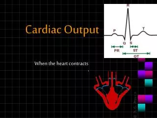
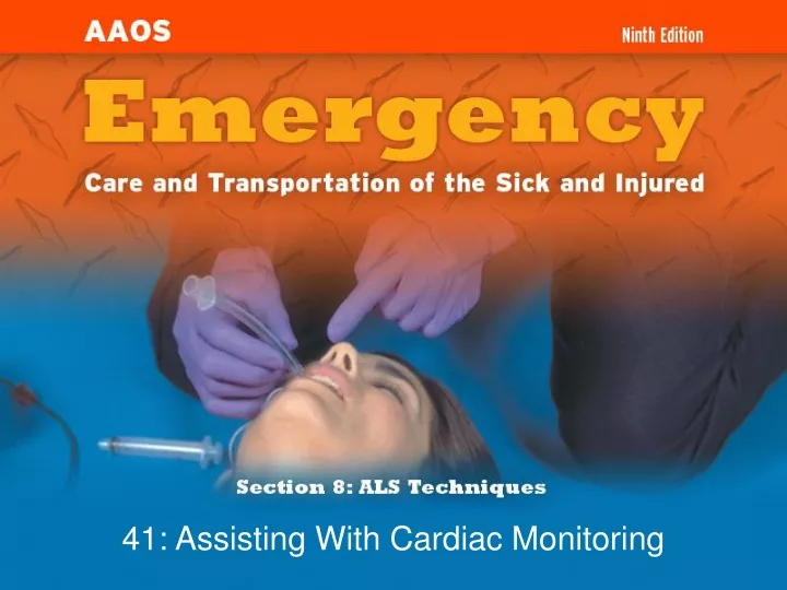
41: Assisting With Cardiac Monitoring. Objectives (1 of 2). To gain an understanding of basic terminology and techniques of cardiac monitoring. To give you the knowledge and tools you need to assist the advanced provider with the use and implementation of an ECG.

E N D
Objectives(1 of 2) • To gain an understanding of basic terminology and techniques of cardiac monitoring. • To give you the knowledge and tools you need to assist the advanced provider with the use and implementation of an ECG. • To better understand the basic anatomy and physiology of the heart.
Objectives (2 of 2) • Identify the components of basic cardiac arrhythmias. • Evaluate the rate and rhythm of a patient’s cardiovascular system, and become familiar with the normal ECG. • Familiarize yourself with and apply 4-lead electrodes and identify placements for the 12-lead systems. *All of the objectives in this chapter are noncurriculum objectives.
Cardiac Monitoring • Use of 12-lead ECGs in the prehospital setting is becoming the norm. • Early identification of AMIs allows hospitals to be prepared. • The EMT-B should know how to place electrodes and leads.
Electrical Conduction System (1 of 2) • A network of specialized tissue in the heart • Conducts electrical current throughout the heart • The flow of electrical current causes contractions that produce pumping of blood.
The Process of Electrical Conduction • Electrical conduction occurs through a pathway of special cells. • SA node: the heart’s main pacemaker • Paces at a rate of 60–100 beats/min • Average of 70 beats/min
Electrodes and Waves Electrodes pick up electrical activity of the heart.
The ECG Complex • One complex represents one beat in the heart. • Complex consists of P, QRS, and T waves.
ECG Paper • Each small box on the paper represents 0.04 seconds. • Five small boxes in larger box represents 0.20 seconds. • Five large boxes equal 1 second.
Normal Sinus Rhythm • Consistent P waves • Consistent P-R interval • 60–100 beats/min
Sinus Bradycardia • Consistent P waves • Consistent P-R interval • Less than 60 beats/min
Sinus Tachycardia • Consistent P waves • Consistent P-R interval • More than 100 beats/min
Ventricular Tachycardia • Three or more ventricular complexes in a row • More than 100 beats/min
Ventricular Fibrillation • Rapid, completely disorganized rhythm • Deadly arrhythmia that requires immediate treatment
Asystole • Complete absence of electrical cardiac activity • Patient is clinically dead. • Decision to terminate resuscitation efforts depends on local protocol.
Cardiac Monitors • May be 3-, 4-, or 12-lead system • Compact, light, portable • Many monitors now combine functions beyond ECG.
12-Lead ECG • Used to identify possible myocardial ischemia • Studies show 12-lead acquisition takes little extra time. • Early identification of acute ischemia and accurate identification of arrhythmias
Lead Placement • EMT-Bs can help in efficient cardiac monitoring by placing electrodes. • Electrodes can be placed while ALS provider prepares other parts of call.
4-Lead Placement • Four leads are called limb leads. • Leads must be placed at least 10 cm from heart.
12-Lead Placement • Limbs leads placed at least 10 cm from heart. • Chest leads must be placed exactly.
Troubleshooting • Clean skin. • Use benzoin. • Shave hair.