Download
1 / 96
960 likes | 1k Views
Gene Expression Systems in Prokaryotes and Eukaryotes. Expression studies Expression in Prokaryotes (Bacteria) Expression in Eukaryotes. Gene Expression Systems in Prokaryotes and Eukaryotes. Expression studies: 1. Analyzing Transcription - Northern blot - Micro array

E N D
Gene Expression Systems in Prokaryotes and Eukaryotes • Expression studies • Expression in Prokaryotes (Bacteria) • Expression in Eukaryotes
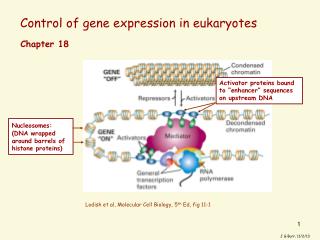
Gene Expression Systems in Prokaryotes and Eukaryotes Expression studies: 1. Analyzing Transcription - Northern blot - Micro array - real-time PCR - Primer extension 2. In vivo Expresion studies Use of report genes to study regulatory elements 3. Analyzing Translation - Western blot - immuno assays - 2D electrophoresis - proteomics
Studying Transcription Microarray technique – DNA chips
Studying Transcription Primer Extension
Promoter Studies • Used reporter genes: • Lac Z • GFP • Luciferase Promoter
Luciferase (luc) systems firefly species Photinus pyralis Expressed luciferase catalyses oxidation of compounds called luciferans ( ATP-dependent process) mouse with a strain of salmonella luciferans emit fluorescense luminometer measurement Mice are injected with LUC+ salmonellas. Sensitive digital cameras allow non-invasive detection. For GT vectors pics look the same
GFP is an extremely stable protein of 238 amino acids with unique post-translationally created and covalently-attached chromophore from oxidised residues 65-67, Ser-Tyr-Gly Green fluorescent protein (GFP) autofluorescent protein from Pacific Northwest jellyfish Aequorea victoria ultraviolet light causes GFP to autofluoresce In a bright green color Jellyfish do nothing with UV, The activate GFP by aequorin (Ca++ activated, biolumuniscent helper)
GFP expression is harmlessfor cells and animals GFP transgenic mice from Osaka University (Masaru Okabe) GFP construct could be used for construct tracking in living organism GFP labelled image of a human tumor. Vessel on the tumor surface are visible in black
Many more fluorescent proteins are engineered San Diego beach scene drawn with living bacteria expressing 8 different colors of fluorescent proteins. Engineered proteins are covering all the spectrum
Use of green fluorescent protein (GFP) as a reporter gene. Page 119
Gene Expression Transcriptional start Translational start
Gene Expression • Gene copy number: • 1. Plasmid copy number: • The copy-number of a plasmid in the cell is determined by regulating the initiation of plasmid replication. • The initiation of plasmid replication may be controlled by: • the amount of available primer (RNA) • the amount of essential replication proteins • the function of essential replication proteins. • 2. Gene dosage -> number of genes integrated into chromosome • - prokaryotic systems -> i.e. Transposons, phages, recombinantion • - mainly eukaryotic systems
Incompatibility of plasmids: Not all plasmids are able to coexist in the same cell. Plasmids which have the same replication control functions are incompatible, and are assigned to the same incompatibility group (inc group). Plasmids of one incompatibility group are related to each other, but cannot survive together in the same bacterial cell, as only different kinds of plasmids are compatible. Ensures that we can make libraries -> just one plasmid taken up by one cell
Homologous integration into chromosome Insertion on Bacillus subtilis chromosome
Protein expression in prokaryotic systems So, this new story would be about vectors again. Bacterial expression vectors have some distinct features: Inducible promoter systems; Protein fusions including fused tags; www.qiagen.com
General advices for one who wants to produce gene expression in prokaryotes Most obvious and common mistakes: 1. Do not forget to cut out the intron 2. Check orientation of insert 3. Do fusions with something In-frame 4. No Post-translation modification = no product activity
Not an issue when you clone a cDNA Introns www.wzw.tum.de/gene-quantification/ mrna.html
Orientation of insert (could go backward, if cloned with same-type sticky ends) – use incompatible sticky ends www.bch.bris.ac.uk/staff/ pfdg/ teaching/genes.htm
Fusion proteins. When expressing a fusion proteins, ensure that both of them are in the same reading frame www.bch.bris.ac.uk/staff/ pfdg/ teaching/genes.htm
PostTranslational modification Eukaryotic cells have Golgi system Prokaryotic cells do not have it nucleus Golgi
Efficiency of expression in E.coli Dependent of: 1. Type of transcription promoter and terminator 2. Affinity of mRNA and prokaryotic ribosome 3. Amount of copies of transgene and its localization (chromosome or plasmid) 4. Cellular localisation of the protein end-product 5. Efficiency of translation in the host organism 6. Stability of protein product in the host organism Systems could be optimized on gene to gene basis. No universal strategy possible
Factors affecting transcription • Promoters (including regulated ones) • PROKARYOTIC!!!! 2. Terminators PROKARYOTIC!!!!
Variations between prokaryotic promoters are minimal http://www.blc.arizona.edu/marty/ 411
Factors affecting translation 1. Ribosome binding site (RBS) 2. Codon bias 3. Stability of the transcript
Ribosome binding site (RBS) =translation initiation site complimentary to 16S rRNA <10 nt Avoid hairpins on 5’ end of gene (minimize GC content) Examining the second codon; better AAA – lysin (13.9% of all E.coli genes). Expression can vary 15 times.
Codon Optimization Strategies • Chemically synthesize new gene • Alter sequence of the gene of interest to match donor codons to the codons most frequently used in host organism • Express in different host • choose host with better matching codon usage • Use an engineered host cell that overexpresses low abundance tRNAs
BL21 (DE3) CodonPlus-RIL (AT-rich compatible) arginine (AGG, AGA), isoleucine (AUA) and leucine (CUA) BL21 (DE3) CodonPlus-RP (GC-rich compatible) arginine (AGG, AGA) and proline (CCC) (AT-rich compatible) Rosetta or Rosetta (DE3) AGG/AGA (arginine), CGG (arginine), AUA (isoleucine) CUA (leucine)CCC (proline), and GGA (glycine) Commercial E. coli strains encode for a number of the rare codon genes
Mitochondria CODON Standard Code: Nuclear-Encoded Proteins Mammals Drosophila Neurospora Yeasts Plants UGA Stop Trp Trp Trp Trp Stop AGA, AGG Arg Stop Ser Arg Arg Arg AUA Ile Met Met Ile Met Ile AUU Ile Met Met Met Met Ile CUU, CUC, CUA, CUG Leu Leu Leu Leu Thr Leu Mitochondria and chloroplast genes Alterations in the Standard Genetic Code in Mitochondria
Factors affecting protein stability • Overall level of protease activity • in bacterial cells 2. N-terminal amino acid affects protein half-life 3. Internal regions containing clusters of certain amino acids can increase proteolysis P prolineE glutamic acidS serineT threonine …. Mutate PEST aminoacids….
Protease-deficient host strains BL21, the work horse of E. coli expression, is deficient in two proteases encoded by the lon (cytoplasmic) and ompT (periplasmic) genes. It is dangerous to kill proteases, it makes E.coli grow much slowly as proteases needed for proper metabolism
Inducible bacterial promoters Why not to use constitutive, always strong promoter? Bacterial grow takes time…. Because recombinant (alien) protein is often toxic for bacterial cell. Bacteria tend to expel harmful plasmids Induction
BL(DE3) inducible system and pET vectors (invented in 1984 by Bill Studier, on sale by Novagen) Gene of interest is expressed from strong T7 promoter pET23 1) T7 RNA polymerase gene is integrated in chromosome under the control of a lac promoter and operator 2) lactose analogue, IPTG, causes the host to produce T7 RNA polymerase • 3) The E. coli host genome also carries the lacI (repressor) gene
Why repressor gene and gene of interest are expressed from different DNA molecules? Repressor gene expressed from chromosome; Gene of Interest expressed from plasmid If too high repressor no transcription (you need to increase expensive IPTG) If too low repressor promoter is leaky (active without IPTG) Repressor is in chromosome, because there it is best kept controlled there (no plasmid loss, not too high expression)
Where your expressed protein will be located? Secreted (!!) E.Coli can not do that Inclusion bodies (insoluble) Cytoplasm (soluble) Periplasmatic space (soluble or insoluble)
1. Inclusion bodies (most common case) -- Inclusion bodies are formed through the accumulation of folding intermediates rather than from the native or unfolded proteins. -- It is not possible to predict which proteins will be produced as inclusion bodies. -- Production of inclusion bodies not dependent on the origin of protein, the used promoters, the hydrophobicity of target proteins...
Electron micrograph of an inclusion body of the protein prochymosin in an E. coli cell Protein Folding Page 116
Good side of inclusion bodies • inclusion bodies can be accumulated in the cytoplasm • to much higher level (greater than 25%) • than production as soluble form; 2) inclusion bodies is initially isolated in a highly purified, solid, and concentrated state by simple physical operation (centrifugation). 3) inclusion bodies have no biological activity. For toxic proteins it may be the only one available; 4) inclusion bodies areresistant to proteolysis That results in the high yield of protein production.
SDS-PAGE analysis of recombinant protein produced as inclusion body hG-CSF mbel.kaist.ac.kr/research/ protein_en1.html
Recovery of proteins from inclusion bodies Is not a straightforward process, but road of trials and errors Refolding Solubilization -- Refolding is initiated by reducing concentration of denaturant used to solubilize IBs. Choice of solubilizing agents, e.g., urea, guanidine HCl, or detergents, plays a key role in solubilization efficiency -- Refolding competes with other reactions, such as misfolding and aggregation (both are leading to bad results) -- Chaperones are helpful in refolding (including chemical chaperones) Guandinium
Diversity of proteins could be exploited Columnchromatography Matrix particles usually packed in the column in the form of small beads. A protein purification strategy might employ in turn each of the three kinds of matrix described below, with a final protein purification Of up to 10,000-fold. Essential Cell Biology: An Introduction to the Molecular Biology of the Cell
Column chromatography Different proteins are retarded to different extents by their interaction with the matrix, they can be collected separately as they flow out from the bottom. According to the choice of matrix, proteins can be separated according to -- their charge, -- their hydrophobicity, -- their size, -- their ability to bind to particular chemical groups (!!) Essential Cell Biology: An Introduction to the Molecular Biology of the Cell
(A) ION-EXCHANGE CHROMATOGRAPHY Ion-exchange columns are packed with small beads that carry positive or negative charges retarding proteins of the opposite charge. The association between a protein and the matrix depends on the pH and ionic strength of the solution passing down the column. These can be varied in a controlled way to achieve an effective separation. Essential Cell Biology: An Introduction to the Molecular Biology of the Cell
(B) GEL-FILTRATION CHROMATOGRAPHY Gel-filtration columns separate proteins according to their size on tiny porous beads. Protein molecules that are small enough to enter the holes in the beads are delayed and travel more slowly through the column. Proteins that cannot enter the beads are washed out of the column first. Such columns also allow an estimate of protein size. Essential Cell Biology: An Introduction to the Molecular Biology of the Cell
(C) AFFINITY CHROMATOGRAPHY Affinity columns contain a matrix covalently coupled to a molecule that interacts specifically with the protein of interest (e.g., an antibody, or an enzyme substrate). Proteins that bind specifically to such a column can finally be released by a pH change or by concentrated salt solutions, and they emerge highly purified. Essential Cell Biology: An Introduction to the Molecular Biology of the Cell