Download
1 / 33
400 likes | 776 Views
Eye and Vision. Exercise 26 BI 232. External Features. Notice the pupil which is surrounded by the colored iris. The sclera is the white of the eye which is covered by a membrane called the conjunctiva Eyelids come together at lateral and medial commissures .

E N D
Eye and Vision Exercise 26 BI 232
External Features • Notice the pupil which is surrounded by the colored iris. • The sclera is the white of the eye which is covered by a membrane called the conjunctiva • Eyelids come together at lateral and medial commissures. • The lacrimalcaruncle is found in the medial commissure.
Lacrimal Apparatus • Consists of the lacrimal gland and its accessory structures. • Produces water, alkaline tears. • Contain antibacterial enzyme called lysozyme for protection from bacterial infections
Extrinsic Eye Muscles • Superior oblique: primarily rotates the top of the eye toward the nose and secondarily moves the eye downward • Innervated by CNIV (trochlear) • Trochlea: Ligament sling • Superior rectus: primarily moves the eye upward and secondarily rotates the top of the eye toward the nose • Innervated by CNIII (oculomotor) • Lateral rectus: moves the eye away from the nose • Innervated by CNVI (abducens)
Extrinsic Eye Muscles • Medial rectus: moves the eye toward the nose • Innervated by CNIII (occulomotor) • Inferior oblique: primarily rotates the top of the eye away from the nose and secondarily moves the eye upward (also CNIII) • Inferior rectus: primarily moves the eye downward and secondarily rotates the top of the eye away from the nose (also CNIII)
Ear Nose
Ciliary body A • Ciliary processes • Ciliary epithelium • Secretes aqueous humor • Ciliary muscle - (intrinsic eye muscle) • Suspensory ligament of the lens A= anterior chamber P= posterior chamber P 3 1 2
Intrinsic Eye Muscles of the Iris • Pupils constrict (Parasympathetic) • Close vision and bright light • Pupils dilate (Sympathetic) • Distant vision and dim light
Neural pathway for vision • After optic nerves exit the eyeballs they meet at the optic chiasm. • Fibers from medial half of retina cross over to the opposite side. • Optic tracts project to the lateral geniculate bodies in thalamus • Some fibers are relayed to superior colliculi
Eye Dissection • Do eye dissection • Be able to ID structures for quizzes and practical
Determination of the Near Point • The minimum distance an object can comfortably be held in focus • Changes as we age • Hold a paper with fine print vertically at arm’s length in front of you. • Close one eye and slowly move the paper closer until either you see two objects or it becomes blurry. • Have partner measure distance
Distribution of Rods and Cones • Slowly move a small colored object, without letting your lab partner see it, from the back of your lab partner’s head, around the side toward the front. • With lab partner still looking straight ahead, not the angle when they are able to see the object.(rods) • Continue to move object forward slowly until color recognition occurs
Binocular Visual Field • Close right eye and look straight ahead with the left • Lab partner moves an object from behind your head from the left until you can see object. • Measure angle with protractor from tip of nose • Continue moving object until it disappears. • Repeat the exercise with the other eye • Determine the total visual field by adding the sums of above.
Visual Acuity • Snellen eye chart used to test visual acuity • 20/20 normal • 20/15 you can see at 20 feet what people with normal vision see at 15 feet. • 20/30 you see at 20 feet what people with normal vision see at 30 feet.
Astigmatism • The degree of curvature in the cornea or lens varies from one axis to another (is uneven or wavy) • This causes light to focus on more than one area of the retina creating a blurry image.
Ophthalmoscope • Used for diagnosis of variances in the eyeball but also as a potential indicator of diseases, such as diabetes mellitus. • Sit facing your lab partner • Have scope set at zero • Move in close to examine the eye of your partner • Look into the pupil
Refraction • Light is bent when it passes from one medium to another medium with a different density • Light passes through these before it hits the retina: • Cornea • Aqueous humor • Lens • Vitreous humor
Focal Point & Focal Distance • Focal Point: The specific point of intersection on the retina. • Focal distance: The distance between the center of the lens and its focal point. Determined by two factors: • Distance from the object to the lens • Shape of the lens
Focal Distance • Distance from the object to the lens: the closer an object is, the greater the focal distance • Shape of the lens: the rounder the lens, the more refraction occurs, so it has a shorter focal distance
Accommodation • Accommodation is an alteration in the curvature of the lens of the eye to focus an image on the retina • Near objects: Lens becomes rounder • Distance objects: Lens becomes flatter
Accommodation • Emmetropia is normal vision. • The image will be focused on the retina’s surface
Accommodation Problems • Myopia: Nearsighted • The eyeball is too deep or the curvature of the lens is too great • The focal point is in front of the retina, so distance objects are blurry • Corrected with a diverging lens
Accommodation Problems • Hyperopia: Farsighted • The eyeball is too shallow or the curvature of the lens is too flat • The focal point is behind of the retina, so near objects are blurry • Corrected with a converging lens
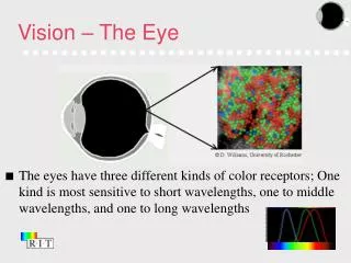
Color Blindness • Cones are responsible for color vision. • 3 types of cones each able to absorb light at specific wavelengths. • Lack of one or more types of cone can cause color blindness • Use Ishihara color plates to test
Afterimages • Photosensitive pigment of the rods is rhodopsin, a light-sensitive retinal and the protein opsin. • When light strikes the retina, rhodopsin splits into its two component parts and becomes pale (bleaching) • You can test the time for separation and reassembly of the photopigments by staring at a contrasting image on a card • Stare about 10-20 seconds and then shuts your eyes. Have your partner record time. You should see colored image against dark background (positive afterimage) After a few moments you should see the reverse of the original image (negative afterimage)
Blind Spot • The optic disc is a region where the nerve fibers exit the back of the eye and form the optic nerve. • This region is devoid of photoreceptors and is called the blind spot. • Follow the instructions from your instructor or book to find your blind spot
The End • Spend the rest of the lab locating structures on the eye models • Make sure that you understand the tests we did in class because you will be asked about them on quizzes and practicals