Download
1 / 31
340 likes | 637 Views
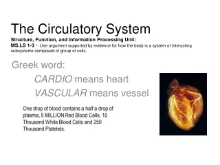
ISSN: 2155-9538. CURRENT RESEARCH ON CARDIO VASCULAR AUTONOMIC FUNCTION TEST. Journal of Bioengineering and Biomedical Sciences. DR.M.RAJAJEYAKUMAR.MBBS,MD(JIPMER),MSc(YOGA),CCEBDM (PHFI) PhD̅ ASSISTANT PROFESSOR, DEPARTMENT OF PHYSIOLOGY,

E N D
ISSN: 2155-9538 CURRENT RESEARCH ON CARDIO VASCULAR AUTONOMIC FUNCTION TEST Journal of Bioengineering and Biomedical Sciences DR.M.RAJAJEYAKUMAR.MBBS,MD(JIPMER),MSc(YOGA),CCEBDM (PHFI) PhD̅ ASSISTANT PROFESSOR, DEPARTMENT OF PHYSIOLOGY, CHENNAI MEDICAL COLLEGE HOSPITAL & RESEARCH CENTRE(SRM GROUP), TRICHY-621105, TAMILNADU, INDIA. .
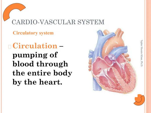
Introduction • Simple bedside tests of cardiovascular autonomic function (Ewing’s tests) have been developed for clinical evaluation of patients suspected to have generalized autonomic dysfunction. ANS PSNS SNS
Indications for AFT • Primary autonomic failure • Secondary autonomic failure 1.DM 2.HT 3.Alcoholism 4.Others
Procedure • The tests were carried out in the Polygraph laboratory of the Department of Physiology between 9.30 am and 12.30 pm, 1-3 hr after a light breakfast. • The laboratory environment was quiet, the temperature between 30 – 35 degrees Celsius and the lighting subdued. • Subjects were asked to empty their bladder before the tests. The tests did not involve intravascular instrumentation or administration of drugs at any stage.
Cardiovascular autonomic function tests • After 10 minutes adaptation in the laboratory,BP was measured in the lying position using a mercury sphygmomanometer. • For a detailed account of the methods of autonomic testing, given in consult Mathias CJ and Bannister R (1992). • During each of the following tests, a bipolar chest lead ECG was recorded continuously and BP was measured using a noninvasive automated BP monitor.
Cardiovascular autonomic function tests The following tests were then performed in the order mentioned below. 1. Baseline BP, HR and HRV in the supine position. (SVB) 2. BP, HR and HRV during standing. (BOTH) 3. Heart rate variation during deep breathing. (PS) 4. BP, HR during sustained isometric handgrip.(SYM) • BP, HR during immersion of right hand in cold water.(SYM) • HR, BP response to Valsalva maneuver.(BOTH)
Cardiovascular autonomic function tests • Baseline BP, HR and HRV: ECG was recorded for at least 330 seconds to determine resting heart rate variability. Baseline BP was recorded in the left arm after 10 minutes of rest in the supine position. HR > 100, BP- >140/90 is abnormal. 2. BP, HR and HRV response to standing: The subject was asked to stand for at least 330 seconds. BP and HR were recorded immediately, 2 minutes and 5 minutes after the standing position.
Cardiac Autonomic Neuropathy Response to standing Autonomic Reflex Arc Blood PressureImmediate standingAbnormal Orthostatic hypotension: Fall in BP 20/10 Heart rate : normally increases 10-20 beats. Heart Rate Record ECG 30 s : 15 s ( RR )< 1.04 This ratio decreases with age.
Cardiovascular autonomic function tests 3. Heart rate variation during deep breathing: With the subject lying down comfortably on a couch, we instructed him to breathe slowly and deeply (following my count – “Breathe in, 1 – 2 – 3 – 4 – 5, breathe out, – 1 – 2 –3 – 4 – 5 and so on) at about six breaths per minute, 5 seconds each for inspiration and expiration. RSA: The maximum of six HR differences (maximum HR minus minimum HR during a deep breathing cycle) was taken as the deep breathing difference.
Cardiac Autonomic Neuropathy RR - Variation during deep breathing Parasympathetic Influence on heart • Maximum- Minimum heart rate < 10 beats /min (Abnormal) • Ratio of longest RR interval ( Expiration ) : Shortest • RR interval ( Inspiration ) { E:I} > 1.17 (Abnormal)
Cardiovascular autonomic function tests 5. BP and HR changes during sustained isometric handgrip: • we asked the subject to maintain 30% of maximum voluntary contraction (MVC) for at least 60 seconds. • BP was monitored in the non-exercising arm after 1 minute of sustained handgrip. After that we asked the subject to discontinue the exercise and recorded BP at once. • Normal response increase DBP > 15mm of Hg and HR (30%).
Cardiovascular autonomic function tests 4. BP and HR changes during immersion of right hand in cold water: • The cold pressor test was performed by having the comfortably seated subject immerse his right hand in cold water at 4 degrees Celsius and then measuring BP after 1 minute of immersion and immediately after taking the hand out of the water. • Response is increased BP >20/10 for normal subjects. • This test is not consistent in all subjects.
Cardiovascular autonomic function tests 6.HR, BP response to Valsalva maneuver: • Forced expiration against open glottis. • Exhale forcefully in to the manometer with close the nostrils with nose clips and maintain the pressure at 40mm of Hg for 10 to 15 sec. • Record the ECG 30 sec before and after the procedure.
Cardiac Autonomic Neuropathy • Phase I Rise in BP, HR. • Phase II in BP ; Tachycardia.(Shortest RR) • Phase III Fall in BP • Phase IV Overshoot of BP; Bradycardia.(longest RR) Valsalva Ratio Longest RR : Shortest RR < 1.2. In normal persons as age increases the VR ratio will decrease. TR= Shortest RR interval during the procedure: Shortest RR interval before the maneuver. BR = Longest RR interval during the procedure: longest RR interval before the maneuver.
Heart rate variability analysis • Heart rate variability has come to be widely used as a noninvasive tool to assess autonomic function in a variety of physiologic as well as disease states. • A detailed account of techniques of heart rate variability analysis is mentioned in the Task force report of the European Society of Cardiology, 1996.
Techniques of heart rate variability analysis: • Briefly, ECG was acquired at a rate of 1000 samples per second using the BIOPAC MP 100 system (BIOPAC Inc., USA) and the BIOPAC AcqKnowledge software 3.7.1 (BIOPAC Inc., USA) for at least 330 seconds during supine rest. • To check the ECG for artifacts and ectopic then edited them out and joined the preceding and successive noise-free segments by linear interpolation with NN intervals (i.e. normal-to-normal RR intervals).
Techniques of heart rate variability analysis • The edited ECG was processed using an R-wave detector to obtain an RR interval tachogram. • Heart rate variability analysis by time- and frequency-domain methods was done using the AcqKnowledge 3.7.1 software (BIOPAC Inc., USA).
References 1. Berntson GG, Bigger JT, Eckberg DL, Grossman P, Kaufmann PG, Malik M, Nagaraja HN, Porges SW, Saul JP, Stone PH, van der Molen MW (1997) Heart rate variability: origins, methods, and interpretivecaveats.BPsychophysiology34: 623-648. 2.Ewing DJ (1992) Analysis of heart rate variability and other non-invasive tests with special reference to diabetic mellitus. In: Bannister R, Mathias CJ, (eds) Autonomic failure. A textbook of clinical disorders of the autonomic nervous system. 3rd edition. Oxford University Press, NY, p 312-333. 3. Task Force. Heart rate variability: Standards of measurement, physiological interpretation and clinical use. Task Force of the European Society of Cardiology and the North American Society of Pacing and Electrophysiology. Circulation 1996; 93:1043–1065.
OMICS Group OMICS Group International through its Open Access Initiative is committed to make genuine and reliable contributions to the scientific community. OMICS Group hosts over 400 leading-edge peer reviewed Open Access Journals and organizes over 300 International Conferences annually all over the world. OMICS Publishing Group journals have over 3 million readers and the fame and success of the same can be attributed to the strong editorial board which contains over 30000 eminent personalities that ensure a rapid, quality and quick review process. OMICS Group signed an agreement with more than 1000 International Societies to make healthcare information Open Access. Contact us at: contact.omics@omicsonline.org
OMICS Group Open Access Membership OMICS publishing Group Open Access Membership enables academic and research institutions, funders and corporations to actively encourage open access in scholarly communication and the dissemination of research published by their authors. For more details and benefits, click on the link below: http://omicsonline.org/membership.php

![CARDIO-VASCULAR SYSTEM [CVS] FUNCTIONAL ANATOMY OF HEART](https://cdn1.slideserve.com/1739818/cardio-vascular-system-cvs-functional-anatomy-of-heart-dt.jpg)