Download
1 / 51
550 likes | 842 Views
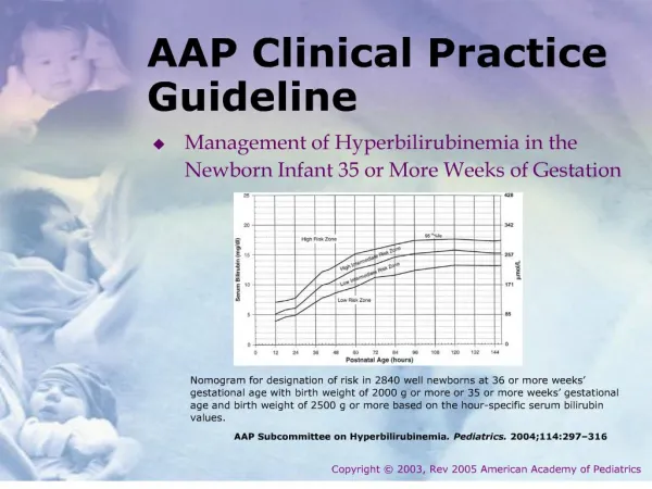
February 2014. Clinical practice guideline on diagnosis and treatment of hyponatraemia. Hierarchy of Outcomes. Formulating statements and grading recommendations. Method of rating the quality of the evidence.

E N D
February 2014 Clinical practice guideline on diagnosisand treatment of hyponatraemia
Hierarchy of Outcomes Formulating statements and grading recommendations
Method of rating the quality of the evidence Method of rating the quality of the evidence. Adapted from Balshem H, Helfand M, Schu¨ nemann HJ, Oxman AD, Kunz R, Brozek J, Vist GE, Falck-Ytter Y, Meerpohl J, Norris S, et al. GRADE guidelines: 3. Rating the quality of evidence. Journal of Clinical Epidemiology 2011 64 401–406. Formulating statements and grading recommendations
Grade of the overall quality of evidence Grade for the overall quality of evidence. Adapted from Guyatt GH, Oxman AD, Vist GE, Kunz R, Falck-Ytter Y, Alonso-Coello P, Schu¨ nemann HJ & GRADE Working Group. GRADE: an emerging consensus on rating quality of evidence and strength of recommendations. BMJ 2008 336 924–926 Formulating statements and grading recommendations
Implications of strong and weak recommendations for stakeholders Implications of strong and weak recommendations for stakeholders. Adapted from Guyatt GH, Oxman AD, Kunz R, Falck- Ytter Y, Vist GE, Liberati A, Schunemann HJ & GRADE Working Group. Going from evidence to recommendations. BMJ 2008 3361049–1051. The additional category ‘Not Graded’ was used, typically, to provide guidance based on common sense or where thetopic does not allow adequate application of evidence. The most common examples include recommendations regardingmonitoring intervals, counselling and referral to other clinical specialists. The ungraded recommendations are generally written assimple declarative statements but are not meant to be interpreted as being stronger recommendations than level 1 or 2 recommendations. Formulating statements and grading recommendations
Grade system for grading recommendations Grade system for grading recommendations. Adapted from Guyatt GH, Oxman AD, Vist GE, Kunz R, Falck-Ytter Y, Alonso-Coello P, Schunemann HJ & GRADE Working Group. GRADE: an emerging consensus on rating quality of evidence and strength of recommendations. BMJ 2008 336 924–926. Formulating statements and grading recommendations
6. Diagnosis of Hyponatraemia 6.1. Classification of hyponatraemia 6.1. Classification of hyponatraemia
6.1.1. Definition of hyponatraemia based on biochemical severity 6.1.1.1. We define ‘mild’ hyponatraemia as a biochemical finding of a serum sodium concentration between 130 and 135 mmol/l as measured by ion-specific electrode. 6.1.1.2. We define ‘moderate’ hyponatraemia as a biochemical finding of a serum sodium concentration between 125 and 129 mmol/l as measured by ionspecific electrode. 6.1.1.3. We define ‘profound’ hyponatraemia as a biochemical finding of a serum sodium concentration <125 mmol/l as measured by ion-specific electrode 6.1. Classification of hyponatraemia
6.1.2. Definition of hyponatraemia based on time of development 6.1.2.1. We define ‘acute’ hyponatraemia as hyponatraemia that is documented to exist <48 h. 6.1.2.2. We define ‘chronic’ hyponatraemia as hyponatraemiathat is documented to exist for at least 48 h. 6.1.2.3. If hyponatraemia cannot be classified, we consider it being chronic, unless there is clinical or anamnestic evidence of the contrary (Table 8). 6.1. Classification of hyponatraemia
Drugs and conditions associated with acute hyponatraemia (<48 h) 6.1. Classification of hyponatraemia
6.1.3. Definition of hyponatraemia based on symptoms 6.1.3.1. We define ‘moderately symptomatic’ hyponatraemia as any biochemical degree of hyponatraemia in the presence of moderately severe symptoms of hyponatraemia (Table 5). 6.1.3.2. We define ‘severely symptomatic’ hyponatraemia as any biochemical degree of hyponatraemia in the presence of severe symptoms of hyponatraemia (Table 5). 6.1. Classification of hyponatraemia
6.2. Confirming hypotonic and excluding non-hypotonic hyponatraemia
6.2. Confirming hypotonic and excluding non-hypotonic hyponatraemia 6.2.1.1. We recommend excluding hyperglycaemic hyponatraemia by measuring the serum glucose concentration and correcting the measured serum sodium concentration for the serum glucose concentration if the latter is increased (1D). 6.2. Confirming hypotonic and excluding non-hypotonic hyponatraemia
6.2. Confirming hypotonic and excluding non-hypotonic hyponatraemia 6.2.1.2. Hyponatraemia with a measured osmolality <275 mOsm/kg always reflects hypotonic yponatraemia (not graded). 6.2.1.3. Accept as ‘hypotonic hyponatraemia’ a hyponatraemia without evidence for causes of non-hypotonic hyponatraemia as listed in Table 10 (not graded). 6.2. Confirming hypotonic and excluding non-hypotonic hyponatraemia
6.3. Which parameters to be used for differentiating causes of hypotonic hyponatraemia?
6.3. Which parameters to be used for differentiating causes of hypotonic hyponatraemia? 6.3.1.1. We recommend interpreting urine osmolality of a spot urine sample as a first step (1D). 6.3.1.2. If urine osmolality is %100 mOsm/kg, we recommend accepting relative excess water intake as a cause of the hypotonic hyponatraemia (1D). 6.3.1.3. If urine osmolality is O100 mOsm/kg, we recommend interpreting the urine sodium concentration on a spot urine sample taken simultaneously with a blood sample (1D). 6.3. Which parameters to be used for differentiating causes of hypotonic hyponatraemia?
6.3. Which parameters to be used for differentiating causes of hypotonic hyponatraemia? 6.3.1.4. If urine sodium concentration is %30 mmol/l, we suggest accepting low effective arterial volume as a cause of the hypotonic hyponatraemia (2D). 6.3.1.5. If urine sodium concentration O30 mmol/l, we suggest assessing extracellular fluid status and use of diuretics to further differentiate likely causes of hyponatraemia (2D). 6.3.1.6. We suggest against measuring vasopressin for confirming the diagnosis of SIADH (2D). 6.3. Which parameters to be used for differentiating causes of hypotonic hyponatraemia?
Table 6| Diagnostic criteria for the syndrome of inappropriate antidiuresis Adapted from Schwartz WB et al. Am J Med 1957; 23: 529-543. [29] and Janicic N et al. Endocrinol Metab Clin North Am 2003; 32: 459-481. [244]
Table 7| Causes of the syndrome of inappropriate antidiuresis
Table 11| Differences between SIADH and cerebral salt wasting Adapted from Sherlock M et al. Clin Endocrinol 2006; 64: 250-254 [42] .
7. Treatment of Hypotonic Hyponatraemia 7.Treatment of Hypotonic Hyponatraemia
7.1. Hyponatraemia with severe symptoms 7.Treatment of Hypotonic Hyponatraemia
7.1.1. First-hour management, regardless of whether hyponatraemia is acute or chronic 7.1.1.1. We recommend prompt i.v. infusion of 150 ml 3% hypertonic for 20 min (1D). 7.1.1.2. We suggest checking the serum sodium concentration after 20 min while repeating an infusion of 150 ml 3% hypertonic saline for the next 20 min (2D). 7.1.1.3. We suggest repeating therapeutic recommendations 7.1.1.1 and 7.1.1.2 twice or until a target of 5 mmol/l increase in serum sodium concentration is achieved (2D). 7.Treatment of Hypotonic Hyponatraemia
7.1.1. First-hour management, regardless of whether hyponatraemiais acute or chronic 7.1.1.4. Manage patients with severely symptomatic hyponatraemia in an environment where close biochemical and clinical monitoring can be provided (not graded). 7.Treatment of Hypotonic Hyponatraemia
7.1.2. Follow-up management in case of improvement of symptoms after a 5 mmol/l increase in serum sodium concentration in the first hour, regardless of whether hyponatraemia is acute or chronic 7.1.2.1. We recommend stopping the infusion of hypertonic saline (1D). 7.1.2.2. We recommend keeping the i.v. line open by infusing the smallest feasible volume of 0.9% saline until cause-specific treatment is started (1D). 7.1.2.3. We recommend starting a diagnosis-specific treatment if available, aiming at least to stabilise sodium concentration (1D). 7.Treatment of Hypotonic Hyponatraemia
7.1.2. Follow-up management in case of improvement of symptoms after a 5 mmol/l increase in serum sodium concentration in the first hour, regardless of whether hyponatraemia is acute or chronic 7.1.2.4. We recommend limiting the increase in serum sodium concentration to a total of 10 mmol/l during the first 24 h and an additional 8 mmol/l during every 24 h thereafter until the serum sodium concentration reaches 130 mmol/l (1D). 7.1.2.5. We suggest checking the serum sodium concentration after 6 and 12 h and daily afterwards until the serum sodium concentration has stabilised under stable treatment (2D). 7.Treatment of Hypotonic Hyponatraemia
7.1.3. Follow-up management in case of no improvement of symptoms after a 5 mmol/l increase in serum sodium concentration in the first hour, regardless of whether hyponatraemia is acute or chronic. 7.1.3.1. We recommend continuing an i.v. infusion of 3% hypertonic saline or equivalent aiming for an additional 1 mmol/l per h increase in serum sodium concentration (1D). 7.1.3.2. We recommend stopping the infusion of 3% hypertonic saline or equivalent when the symptoms improve, the serum sodium concentration increases 10 mmol/l in total or the serum sodium concentration reaches 130 mmol/l, whichever occurs first (1D). 7.Treatment of Hypotonic Hyponatraemia
7.1.3. Follow-up management in case of no improvement of symptoms after a 5 mmol/l increase in serum sodium concentration in the first hour, regardless of whether hyponatraemia is acute or chronic. 7.1.3.3. We recommend additional diagnostic exploration for other causes of the symptoms than hyponatraemia (1D). 7.1.3.4. We suggest checking the serum sodium concentration every 4 h as long as an i.v. infusion of 3% hypertonic saline or equivalent is continued (2D). 7.Treatment of Hypotonic Hyponatraemia
7.2. Hyponatraemia with moderately severe symptoms 7.Treatment of Hypotonic Hyponatraemia
7.2. Hyponatraemia with moderately severe symptoms 7.2.1.1. We recommend starting prompt diagnostic assessment (1D). 7.2.1.2. Stop, if possible, medications and other factors that can contribute to or provoke hyponatraemia (not graded). 7.2.1.3. We recommend cause-specific treatment (1D). 7.2.1.4. We suggest immediate treatment with a single i.v. infusion of 150 ml 3% hypertonic saline or equivalent over 20 min (2D). 7.Treatment of Hypotonic Hyponatraemia
7.2. Hyponatraemia with moderately severe symptoms 7.2.1.5. We suggest aiming for a 5 mmol/l per 24 h increase in serum sodium concentration (2D). 7.2.1.6. We suggest limiting the increase in serum sodium concentration to 10 mmol/l in the first 24 h and 8 mmol/l during every 24 h thereafter, until a serum sodium concentration of 130 mmol/l is reached (2D). 7.2.1.7. We suggest checking the serum sodium concentration after 1, 6 and 12 h (2D). 7.Treatment of Hypotonic Hyponatraemia
7.2. Hyponatraemia with moderately severe symptoms 7.2.1.8. We suggest additional diagnostic exploration for other causes of the symptoms if the symptoms do not improve with an increase in serum sodium concentration (2D). 7.2.1.9. We suggest considering to manage the patient as in severely symptomatic hyponatraemia if the serum sodium concentration further decreases despite treating the underlying diagnosis (2D). 7.Treatment of Hypotonic Hyponatraemia
7.3. Acute hyponatraemia without severe or moderately severe symptoms 7.Treatment of Hypotonic Hyponatraemia
7.3. Acute hyponatraemia without severe or moderately severe symptoms 7.3.1.1. Make sure that the serum sodium concentration has been measured using the same technique used for the previous measurement and that no administrative errors in sample handling have occurred (not graded). 7.3.1.2. If possible, stop fluids, medications and other factors that can contribute to or provoke hyponatraemia (not graded). 7.3.1.3. We recommend starting prompt diagnostic assessment (1D). 7.Treatment of Hypotonic Hyponatraemia
7.3. Acute hyponatraemia without severe or moderately severe symptoms 7.3.1.4. We recommend cause-specific treatment (1D). 7.3.1.5. If the acute decrease in serum sodium concentration exceeds 10 mmol/l, we suggest a single i.v. Infusion of 150 ml 3% hypertonic saline or equivalent over 20 min (2D). 7.3.1.6. We suggest checking the serum sodium concentration after 4 h, using the same technique as used for the previous measurement (2D). 7.Treatment of Hypotonic Hyponatraemia
7.4. Chronic hyponatraemia without severe or moderately severe symptoms 7.Treatment of Hypotonic Hyponatraemia
7.4. Chronic hyponatraemia without severe or moderately severe symptoms 7.4.1. General management 7.4.1.1. Stop non-essential fluids, medications and other factors that can contribute to or provoke hyponatraemia (not graded). 7.4.1.2. We recommend cause-specific treatment (1D). 7.4.1.3. In mild hyponatraemia, we suggest against treatment with the sole aim of increasing the serum sodium concentration (2C). 7.Treatment of Hypotonic Hyponatraemia
7.4. Chronic hyponatraemia without severe or moderately severe symptoms 7.4.1. General management 7.4.1.4. In moderate or profound hyponatraemia, we recommend avoiding an increase in serum sodium concentration of O10 mmol/l during the first 24 h and O8 mmol/l during every 24 h thereafter (1D). 7.4.1.5. In moderate or profound hyponatraemia, we suggest checking the serum sodium concentration every 6 h until the serum sodium concentration has stabilised under stable treatment (2D). 7.4.1.6. In case of unresolved hyponatraemia, reconsider the diagnostic algorithm and ask for expert advice (not graded). 7.Treatment of Hypotonic Hyponatraemia
7.4. Chronic hyponatraemia without severe or moderately severe symptoms 7.4.2. Patients with expanded extracellular fluid 7.4.2.1. We recommend against a treatment with the sole aim of increasing the serum sodium concentration in mild or moderate hyponatraemia (1C). 7.4.2.2. We suggest fluid restriction to prevent further fluid overload (2D). 7.4.2.3. We recommend against vasopressin receptor antagonists (1C). 7.4.2.4. We recommend against demeclocycline (1D). 7.Treatment of Hypotonic Hyponatraemia
7.4. Chronic hyponatraemia without severe or moderately severe symptoms 7.4.3. Patients with SIAD 7.4.3.1. In moderate or profound hyponatraemia, we suggest restricting fluid intake as first-line treatment (2D). 7.4.3.2. In moderate or profound hyponatraemia, we suggest the following can be considered equal secondline treatments: increasing solute intake with 0.25–0.50 g/kg per day of urea or a combination of low-dose loop diuretics and oral sodiumchloride (2D). 7.4.3.3. In moderate or profound hyponatraemia, we recommend against lithiumordemeclocycline (1D). 7.Treatment of Hypotonic Hyponatraemia
7.4. Chronic hyponatraemia without severe or moderately severe symptoms 7.4.3. Patients with SIAD 7.4.3.4. In moderate hyponatraemia, we do not recommend vasopressin receptor antagonists (1C). 7.4.3.5. In profound hyponatraemia, we recommend against vasopressin receptor antagonists (1C). 7.Treatment of Hypotonic Hyponatraemia
7.4. Chronic hyponatraemia without severe or moderately severe symptoms 7.4.4. Patients with reduced circulating volume 7.4.4.1. We recommend restoring extracellular volume with i.v. infusion of 0.9% saline or a balanced crystalloid solution at 0.5–1.0 ml/kg per h (1B). 7.4.4.2. Manage patients with haemodynamic instability in an environment where close biochemical and clinical monitoring can be provided (not graded). 7.4.4.3. In case of haemodynamic instability, the need for rapid fluid resuscitation overrides the risk of an overly rapid increase in serum sodium concentration (not graded). 7.Treatment of Hypotonic Hyponatraemia
7.5. What to do if hyponatraemia is corrected too rapidly? 7.5.1.1. We recommend prompt intervention for re-lowering the serum sodium concentration if it increases O10 mmol/l during the first 24 h or O8 mmol/l in any 24 h thereafter (1D). 7.5.1.2. We recommend discontinuing the ongoing active treatment (1D). 7.5.1.3. We recommend consulting an expert to discuss if it is appropriate to start an infusion of 10 ml/kg body weight of electrolyte-free water (e.g. Glucose solutions) over 1 h under strict monitoring of urine output and fluid balance (1D). 7.Treatment of Hypotonic Hyponatraemia
7.5. What to do if hyponatraemia is corrected too rapidly? 7.5.1.4. We recommend consulting an expert to discuss if it is appropriate to add i.v. desmopressin 2 mg, with the understanding that this should not be repeated more frequently than every 8 h (1D). 7.Treatment of Hypotonic Hyponatraemia
Figure 2| Adaptation of the brain to hypotonicity Reproduced with permission from Adrogué HJ et al. New Engl J Med 2000; 342: 1581-1589.
Figure 3| Osmotic stimulation of vasopressin release Schematic representation of normal physiological relationships among plasma osmolality, plasma AVP concentrations, urine osmolality, and urine volume in man. Note particularly the inverse of nature of the relation between urine osmolality and urine volume, resulting in disproportionate effects of small changes in plasma AVP concentrations on urine volume at lower AVP levels. Reproduced with permission from Verbalis JG. Best Prac Res En 2003; 17: 471-503.
Figure 4| Effects of hypovolaemia on osmoreceptor gain Reproduced with permission from Robertson GL et al. J ClinEndocrMetab 1976; 42: 613-620.
Figure 5| Pseudohyponatraemia Normally, serum contains 7 percent solids by volume. In order to reduce the volume of blood needed for analysis, serum is frequently diluted before the actual measurement is obtained. The same volume of diluent is always used; the degree of dilution is estimated under the assumption that the serum contains 7 percent solid-phase particles. When the fraction of solid-phase particles is increased, the same amount of diluent results in a greater dilution, unbeknownst to the laboratory personnel (right side of figure). Consequently, the calculation of an ion level with the use of a degree of dilution that is based on the incorrect fraction of solid-phase particles will lead to an underestimate. Reproduced with permission from Turchin A et al. NEJM 2003; 349: 1465-1469. [92]
Diagnostic Flowchart
Management flowchart