Download
1 / 77
960 likes | 1.39k Views
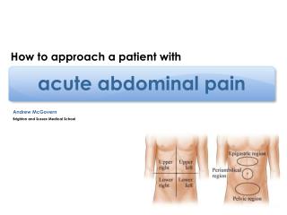
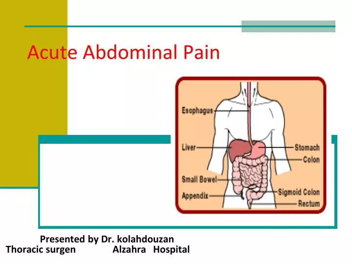
Acute Abdominal Pain. Presented by Dr. kolahdouzan Thoracic surgen Alzahra Hospital. Acute Abdomen. Challenge to Surgeons & Physicians Most common cause of surgical emergency admission Encompass various conditions ranging from the trivial to the life-threatening

E N D
Acute Abdominal Pain Presented by Dr. kolahdouzan Thoracic surgen Alzahra Hospital
Acute Abdomen • Challenge to Surgeons & Physicians • Most common cause of surgical emergency admission • Encompass various conditions ranging from the trivial to the life-threatening • Clinical course can vary from minutes to hours, to weeks • It can be an acute exacerbation of a chronic problem e.g. Chronic Pancreatitis, Vascular Insufficiency
Outline – Acute Abdominal Pain • Intra-abdominal Diagnosis by Organ System : • Gastrointestinal Gynecologic Pain • Appendicitis Acute PID • Biliary Tract Disease Ectopic Preg • Small Ball Obstruction • Diverticulitis Vascular • Acute Pancreatitis AAA • Genitourinary Mesenteric Ischemia • Renal Colic Ischemic Colitis • Acute Urinary Retention / UTI • Treatment • Disposition
The primary symptom of the "acute abdomen" is–Abdominal pain.
ASSESMENT • A Full history • Thorough physical examination Diagnosis can be made most of the time by a good history and a proper physical examination. - An exact diagnosis often impossible to make after the initial assessment, and often relying on further investigation
Types of Abdominal Pain: • Three types of pain exist: 1. Visceral 2. Parietal 3. Referred
1. Visceral Pain • Due to stretching of fibers innervating the walls of hollow or solid organs. • It occurs early and poorly localized • It can be due to early ischemia or inflammation.
2. Parietal Pain • Caused by irritation of parietal peritoneum fibers. • It occurs late and better localized. • Can be localized to a dermatome superficial to site of the painful stimulus.
3. Referred Pain • Pain is felt at a site away from the pathological organ. • Pain is usually ipsilateral to the involved organ and is felt midline if pathology is midline. • Pattern based on developmental embryology.
Acute Abdominal Pain • Two approaches to evaluate pts with acute abdominal pain: 1. Classification of abd pain into systems 2. Abdominal Topography (4 quadrants)
Classification on Abdominal Pain • Three main categories of abdominal pain: 1. Intra-abdominal (arising from within the abd cavity / retroperitoneum) involves: • GI (Appendicitis, Diverticulitis, etc, etc, etc) • GU (Renal Colic, etc, etc, etc) • Gyn (Acute PID, Pregnancy, etc) • Vascular systems (AAA, Mesenteric Ischemia, etc)
Classification on Abdominal Pain 2. Extra-abdominal (less common) involves: • Cardiopulmonary (AMI, etc) • Abdominal wall (Hernia, Zoster etc) • Toxic-metabolic (DKA, OD, lead, etc) • Neurogenic pain (Zoster, etc) • Psychic (Anxiety, Depression, etc) 3. Nonspecific Abd pain – not well explained or described.
Abdominal Topography • RUQ LUQ • RLQ LLQ • UPPER ABDOMEN • LOWER ABDOMEN • CENTRAL • GENERALIZED
Historical features of Abd Pain • Location, quality, severity, onset, and duration of pain, aggravating and alleviating factors • GI symptoms (N/V/D) • GU symptoms • Vascular symptoms (A. fib / AMI / AAA) • Can overlap i.e. Nausea seen in both GI / GU pathologies.
Historical features of Abd Pain • PMH • Recent / current medications • Past hospitalizations • Past surgery • Chronic disease • Social history • Occupation / Toxic exposure (CO / lead)
Physical Examination of the Abdomen • Note pt’s general appearance. Realize that the intensity of the abdominal pain may have no relationship to severity of illness. • One of the initial steps of the PE should be obtaining and interpreting the vitals. • Pts with visceral pain are unable to lie still. • Pts with peritonitis like to stay immobile.
Physical Examination of the Abdomen • INSPECT for distention, scars, masses, rash. • AUSCULATE for hyperactive, obstructive, absent, or normal bowel sounds. • PALPATION to look for guarding, rigidity, rebound tenderness, organomegally, or hernias. • Women should have pelvic exam (check FHR if pregnant). • Anyone with a rectum should have rectal exam (If no rectum check the ostomy).
Laboratory Test • CBC (limited clinical utility) • UA / Urine culture • Lactic acid • LFT / Amylase / Lipase • CE / Troponin • HCG (quant / qual) • Stool Culture
Radiographic Test • Plain abdominal radiographs or abdominal series has several limitations and is subject to reader interpretation. • CT scan in conjunction with ultrasound is superior in identifying any abnormality seen on plain film.
Specific Diagnoses • In patients above fifty years of age the top four reasons for acute abdominal pain are: Biliary Tract Disease (21%,) NSAP (16%), Appendicitis(15%), and Bowel Obstruction (12%). • In patients under fifty years of age the top three reasons for acute abdominal pain are: NSAP (40%,) Appendicitis (32%,) and Other (13%.)
Acute Appendicitis • “In spite of a large number of algorithms and decision rules incorporating many different clinical and laboratory features, an accurate preoperative diagnosis of appendicitis has remain elusive for more than a century.”
Acute Appendicitis • Clinical features with some predictive value include: • Pain located in the RLQ • Pain migration from the periumbilical area to the RLQ • Rigidity • Pain before vomiting • Positive psoas sign • Note: Anorexia is not a useful symptom (33% pts not anorectic preoperatively.)
Acute Appendicitis • Ultrasound can be used for detection, but CT is preferred in adults and non-pregnant women. • The CT scan can be with and without contrast (oral & IV.) • A neg. CT does not exclude diagnosis, but a positive scan confirms it.
Biliary Tract Disease • Most common diagnosis in ED of pts > 50. • Composed of: • Acute Cholecystitis (acalculus / calculus) • Biliary Colic • Common Duct Obstruction (Ascending Cholangitis – painful jundice / fever / MSΔ). • Of those patients found to have acute cholecystitis, the majority lack fever and 40% lack leukocytosis.
Biliary Tract Disease • Patients may complain of: • Diffuse pain in upper half of abdomen • Generalized tenderness throughout belly • RUQ or RLQ pain.
Biliary Tract Disease • Sonography (US) is the initial test of choice for patients with suspected biliary tract disease. More sensitive than CT scan to detect CBD obstruction. • CT scan is better in the identification of cholecystitis than in the detection of CBD obstruction. • Cholescintigraphy (radionclide / HIDA scan) of the biliary tree is a more sensitive test than US for the diagnosis of both of these conditions.
Biliary Tract Disease • MR cholangiography (MRCP) • Has good specificity and sensitivity in picking up stones and common duct obstructions. • Less invasive / less complications than ERCP (ERCP can induce GI perforation, pancreatitis, biliary duct injury)
Small Bowel Obstruction • SBO may result from previous abdominal surgeries. • Patient may present with intermittent, colicky pain, abdominal distention, and abnormal BS. • Only 2 historical features (previous abd surgery and intermittent / colicky pain) and 2 physical findings (abd distention and abn BS) appear to have predictive value in diagnosing SBO.
Small Bowel Obstruction • Plain abd films has a large number of indeterminate readings and can be very limited due to the following: • Pt is obese • Pt is bedridden / contracted (limited lateral decub / upright view) • Technical limitations
Bowel Obstruction • Small BowelLarge Bowel • Central Peripheral • Valvulae conniventes Haustrae • Dia > 5cm > 10cm
Small Bowel Obstruction • CT scan is better than plain film in detecting high grade SBO. • CT scan can also give more info that might not be seen on plain film (i.e. ischemic bowel) • Low grade SBO may require small bowel follow through.
Acute Pancreatitis • 80% of cases are due to ETOH abuse or gallstones. • Other common causes: • Drugs ( Valproic acid, Tetracycline, Hydrochlorothiazide, Furosemide) • Pancreatic cancer • Abdominal trauma/surgery • Ulcer with pancreatic involvement • Familial pancreatitis (Hypertriglycerides / Hypercalcemia) • Iatrogenic (ERCP) • In Trinidad, the sting of the scorpion Tityus trinitatis is the most common cause of acute pancreatitis • Definition : • Inflammation of the pancreas • Associated with edema, pancreatic autodigestion, necrosis and possible hemorrhage
Acute Pancreatitis • Only a minority number of pts present with pain and tenderness limited to the anatomic area of the pancrease in the upper half of the abdomen. • 50% of pts present with c/o pain extending well beyond the upper abd to cause generalized tenderness.
Acute Pancreatitis • The inflammatory process around the pancreas may cause other signs and symptoms such as: • Pleural effusion • Grey Turner's sign ( flank discoloration ) • Cullen's sign ( discoloration around the umbilicus ) • Ascites • Jaundice
Acute Pancreatitis • Lipase testing is preferred in ED. • Other test to consider: (CBC, Amylase, UA and CE/trop) • The height of the pancreatic enzyme elevations do not have prognostic value • A double contrast helical CT scan stages severity and predicts mortality sooner than Ranson’s Criteria.
Acute Pancreatitis • Should consider ICU admission for pts with high Ranson’s Criteria. • When making the diagnosis of Acute Pancreatitis, it maybe necessary to assess the pt for the following: • Biliary pancreatitis • Peripancreatic complications
Acute Pancreatitis Biliary pancreatitis -Due to CBD obstruction. -Can lead to Ascending Cholangitis Clinical findings: May have a fever, jaundice / icterus Lab findings: ↑AST / ALT, ↑Total Bilirubin Radiological std: MRCP - Test of choice to get clear images of the pancrease and CBD. Double contrast CT - can also be use, may have limited view of the CBD – 2nd most common test to be ordered in ED Ultrasound – 1st most common test to be order in ED to evaluate for CBD obstruction. More sensitive than CT scan to evaluate the CBD. Its use is safer in pregnancy.
Acute Pancreatitis Peripancreatic complications: • Necrosis (Necrotizing Pancreatitis) • Hemorrhage (Hemorrhagic Pancreatitis) • Drainable fluid collections (Ruptured Pancreatic Pseudocyst) • Clinical findings: May have a distended Abd, appear septic, Cullen’s sign, and / or Grey Turner’s Sign. • Lab findings:No definite lab test will help in the diagnosis. May see decrease Hg or ↑Lactic Acid level. • Radiological test: of choice to evaluate for the above complications is a double contrast CT scan.
Acute Diverticulitis • Less than ¼ of pts present with LLQ pain. • 1/3 of pts present with pain to the lower half of the abdomen. • 20% of elderly pts with operatively confirmed diverticulitis lacked abdominal tenderness. • Elderly pts are at risk for a severe and often fatal complication of diverticulitis. (Free perforation of the colon)
Acute Diverticulitis • CT with contrast: • Test of choice for Acute Diverticulitis. • Can identify abscesses, other complications, and inform surgical management strategies. • US: • Relies on identification of an inflamed diverticulum to make the diagnosis which is often obscured in pts with complicated diverticulitis.
Renal Colic • Pts may present with abrupt, colicky, unilateral flank pain that radiates to the groin, testicle, or labia. • Hematuria and plain abd films can be helpful however do not provide a strong support in the diagnostic evaluation of suspected renal colic. • Noncontrast helical CT is standard for the diagnosis. IVP has poor sensitivity and time consuming in ED setting. • Must rule out AAA.
Acute Pelvic Inflammatory Disease • Patient may complain of pain / tenderness in lower abdomen, adnexal or cervix. • Most importantly patient may complain of abnormal vaginal discharge (most common finding). • Fever, palpable mass, ↑WBC have been inconsistently associated with PID. • The best noninvasive test is transvaginal ultrasound.
Ectopic Pregnancy • Symptoms include abdominal pain (most common) and vaginal bleeding (maybe the only complaint). • Female pts (child bearing age) that present with these symptoms automatically get a pregnancy test and HCG quantitative level.
Ectopic Pregnancy • If the pt is pregnant, then order a transvaginal US to evaluate for ectopic pregnancy. • Clear view of an IUP in 2 perpendicular views essentially excludes an ectopic pregnancy. • If an IUP is not seen, this must be interpreted in the context of the discriminatory zone (DZ) of the quantitative HCG.
Ectopic Pregnancy • The DZ (1500 mlU/ml) is the threshold level of serum HCG, above which a normal IUP should be seen on sonography. • Although there is a broad range of normal variation in HCG, failure of levels to increase by about 66% within 48 h in 1st trim pregnancy suggests an abnormal gestation (either a threatened miscarriage or blighted pregnancy from an ectopic.) • If the diagnosis is not made with US and there is still a high suspicion for ectopic than laparoscopy is indicated.
Abdominal Aortic Aneurysm • Dissections produce chest or upper back pain that can migrates to abdomen as the dissection extend distally. • AAA rather than dissect, it enlarge, leak, and rupture. • <50% of pts with AAA present with hypotension, abdominal/back pain, and/or pulsatile abd mass. Can present similar to renal colic. • Neither the presence or the absence of femoral pulse or an abdominal bruit are helpful clinically.
Abdominal Aortic Aneurysm • Palpation is an important part of physical exam. Maybe able to detect an enlarged aorta. • Any stable pt > 50 yrs old presenting with recent onset of abd / flank / low back pain should have a CT scan to exclude AAA from the differential diagnosis. • Can use bedside ultrasound FAST scan, but this will not provide information about leakage or rupture. • MRI is limited in its ability to identify fresh bleeding. It is not an appropriate emergency procedure.