Download
1 / 59
1.22k likes | 2.25k Views
Normal EEG in children EEG workshop. Sorawit Viravan. Electrode placement. International 10-20 system Minimum 21 electrodes Odd-numbered electrodes are placed on the left side of the head, and even-numbered electrodes are placed on the right side of the head

E N D
Normal EEG in childrenEEG workshop Sorawit Viravan
Electrode placement • International 10-20 system • Minimum 21 electrodes • Odd-numbered electrodes are placed on the left side of the head, and even-numbered electrodes are placed on the right side of the head • Specific letters designate the anatomical area; for example “F” means frontal
Common Montage types The difference in voltage between two electrodes • Bipolar: Each channel represents difference between 2 adjacent electrodes - AP bipolar - Coronal / transverse bipolar • Referential: Each channel represents the difference between a certain electrode and the designated reference position - ipsilateral ear - average - midline, etc.
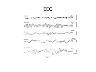
EEG activity • Waveform • Frequency • Amplitude • Polarity • Timing
Spike - Sharply contoured, duration 20-70 msec Sharp wave - Sharply contoured, duration 70-200 msec Sharp transient - Sharply contoured waveform Other morphology - spindles, arciform, saw-tooth Wave form
Wave form • Monophasic wave • Single deflection: up or down • Diphasic wave • 2 components on opposite sides • Polyphasic wave • 2 or more components of different direction
Frequency • Delta wave < 4 Hz • Theta wave 4-7 Hz • Alpha wave 8-13 Hz • Beta wave > 13 Hz
Amplitude Total vertical distance of wave • Low < 20 μV • Medium 20-50 μV • High > 50 μV Affected by barriers
Distribution • Generalized / diffuse • Lateralized • Focal / localized
Polarity Bipolar (input 1 – Input 2) • Upward pen deflection - when input 1 is more negative than input 2 - when input 2 is more positive than input 1 • Downward deflection - when input 1 is more positive than input 2 - when input 2 is more negative than input 1 Referencial (Input 1 – Ref) - negative is up and positive is down
Synchronous Bilaterally synchronous Asynchronous Independent Timing
EEG in newborn • Post conceptional age • Duration at least 60 minutes • Awake / Active sleep / Quiet sleep • Continuity / Synchrony • Symmetry / Reactivity • Normal specific EEG pattern
Awake • Posterior dominant rhythm (PDR) • Posterior slow wave of youth (PSWY) • Mu rhythm • Beta activity • Lambda wave • Eye movement • Artifact
PDR • Alert, eye-closed, in rest state • First seen at 3 months of age • Maximum posterior head region • Reactivity • Attenuation with eye opening, + anxiety
PDR • Higher amplitude over right hemisphere (< 50% difference) due to asymmetric skull thickness • Amplitude ~ 50-100 uV • Decreasing amplitude with increasing age due to increased bone density of the skull
PDR Frequency in Children 3-4 months: 4 Hz 12 months: 5-6 Hz 2 years: 7 Hz 3 years: 8 Hz 9 years: 9 Hz 15 years: 10 Hz
PSWY • Slow activity intermixed with PDR • Moderate voltage (<120% of normal alpha rhythm voltage ) • May be asymmetry • Best seen in 8-14 years • Block with eye opening • Disappear with the alpha rhythm during drowsiness and light sleep
Mu • central arch-like rhythm of alpha frequency (usually 8-10 Hz ) • May be related to the functions of the sensorimotor cortex at rest • Best seen between 8-16 years • Asymmetrical • Blocked unilaterally with movement of the contralateral extremity • Not blocked by eye opening
Beta • Frequencies more than 13 Hz • Amplitude < 20 uV, usually < 10 uV • Three band 18-25 Hz band (common) 14-16 Hz band (less common) 35-40 Hz band (rare) • Increased by • Drugs eg. barbiturate, benzodiazepine, chloral hydrate (18-25 Hz > 14-16 Hz)
Lambda wave • Surface positive, check mark-like wave • Occipital region • During eye opening • Visually scanning at complex picture (ceiling, TV etc.) with saccadic eye movement • Best seen in 2-15 years • May be asymmetrical
Eye movement (EM) Vertical EM (Fp1, Fp2) • Eye opening • Eye closure • Eye blinking Horizontal EM (F7, F8) • To the left • To the right
Eye movement (EM) • Cornea positivity • Retina negativity • Nearest electrode of the direction of EM will pick up positivity, the opposite electrode will pick up negativity
Vertical EM • Eye closure (relatively eyes go up) Fp1 and Fp2 pick up positivity downward deflection at Fp1-F7, Fp2-F8 • Eye opening (relatively eyes go down) Fp1 and Fp2 pick up negativity upward deflection at Fp1-F7, Fp2-F8
Horizontal EM • Eye turn to the left F7 pick up positivity, F8 pick up negativity positive phase reversal at F7 (Hole) negative phase reversal at F8 • Eye turn to the right F8 pick up positivity, F7 pick up negativity positive phase reversal at F8 (Hole) negative phase reversal at F7
Eye to the left + -
Sleep Non-REM sleep • Stage 1 (drowsiness) • Stage 2 • Stage 3 & 4 REM sleep
Stage 1 • Alpha drop out • Hypnagogic hypersynchrony • POSTs • Beta activity • Vertex wave
Hypnagogic hypersynchrony • Burst of generalized high voltage 3-5 Hz • Maximum fronto-central • Awake sleep • Begin 6 months • Best seen 1-5 years • Rare after 11-12 years • Hypnapompic: sleep awake
POSTs • Positive occipital sharp transients of sleep • 4-5 Hz, checkmark-like, isolated or in trains • Esp. daytime nap, arousal return to sleep • Commonly asymmetry • Age 4-50 years • Best seen 15-35 years
Vertex wave • Sharp transient maximum Cz (vertex) • Begin 8 weeks post term • Age 1-4 years; spiky and high amplitude • Runs of vertex
Stage 2 • Sleep spindles • K-complex • Delta wave • (Vertex, POSTs)
Sleep spindles • 11-14 Hz • Maximum central, frontal (Cz, C3C4, F3F4) • 2-5 seconds duration, may be spiky • Lack of fusiform shape as in adult • Begin 6-8 weeks post term; asynchronous but symmetrical • Age 2 years; synchronous
K-complex • Vertex + spindles • Biphasic high amplitude slow wave > 0.5 seconds duration • Maximum Cz (vertex) • Begin 5 months