Download
1 / 21
220 likes | 432 Views
Anatomy of the Urinary System. Kidneys Ureters Bladder Urethra. The Kidneys. Functions of the Kidneys: - removal of waste products from blood - control of blood volume - control of salt balance in blood - control of pH of the blood (acid-base balance).

E N D
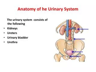
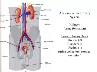
Anatomy of the Urinary System Kidneys Ureters Bladder Urethra
The Kidneys • Functions of the Kidneys: - removal of waste products from blood - control of blood volume - control of salt balance in blood - control of pH of the blood (acid-base balance)
Gross Anatomy of the Kidneys • The kidneys are bean-shaped organs located on the posterior abdominal wall
Gross Anatomy of the Kidneys • Kidneys are retroperitoneal • Kidneys are covered by a renal capsule, and enclosed in a renal fat pad • The kidney and associated fat pad are enclosed by renal fascia
renal pyramids cortex medulla major calyx renal pelvis minor calyx hilus renal columns ureter renal papilla renal capsule Gross Anatomy of the Kidneys • Renal hilus: site where renal artery enters and renal vein and ureter leave the kidney • Internal structures: - cortex - medulla (pyramids) - renal columns - renal papillae - minor calyx - major calyx - renal pelvis -ureter
Renal Blood Supply • Blood goes to the kidney in the renal artery, which branches into segmental arteries as it enters the hilus • Segmental arteries branch into interlobar arteries, which become arcuate arteries as they curve over the base of the renal pyramids • Arcuate arteries give off interlobular arteries in the cortex, from which branch the afferent arterioles (to the nephron) • Efferent arterioles carry blood away from the nephron, into the peritubular capillary network • The capillaries drain into interlobular veins, which drain into arcuate, interlobar, and renal veins
segmental artery interlobar arteries renal artery interlobular arteries renal vein interlobar veins arcuate veins arcuate arteries Renal Blood Supply interlobular artery arcuate artery interlobar vein interlobar artery
proximal convuluted tubule renal corpuscle distal convuluted tubule collecting duct descending limb ascending limb Histology of the Kidney • The basic functional unit of the kidney is the nephron • The nephron is composed of: - renal corpuscle - Bowman’s capsule - glomerulus (capillary tuft) - proximal convuluted tubule - Loop of Henle - distal convuluted tubule • Each nephron is connected to a collecting duct
Location of Functional Units • Nephrons are located primarily in the cortex; Loops of Henle extend into medullary pyramids • Juxtamedullary nephrons are located near medulla, have longer Loops of Henle • Collecting ducts are found in the medullary pyramids, and empty urine into the renal papillae
parietal epithelium efferent arteriole podocyte DCT macula densa glomerular capillary juxtaglomerular cells afferent arteriole Renal Corpuscle • The renal corpuscle (site of filtration) is composed of Bowman’s capsule and the glomerulus • Bowman’s capsule is double-walled structure • Podocyte cells • glomerulus: capillary tufts • gaps in epithelium: fenestrae • juxtaglomerular cells: renin • macula densa
The Ureters • Function: carry urine from the kidneys to the bladder • Histology: three layers to ureter wall - mucosa: transitional epithelium, lamina propria - muscularis: inner longitudinal, outer circular, outermost longitudinal in lowest 1/3rd - adventitia • Enter bladder as narrow slit-like opening
The Bladder • Function: storage of urine • Located in pelvic cavity, posterior to the pubic symphysis • Histology: mucosa, muscularis, and adventitia, as in ureters (thicker muscularis in bladder) • Specialization of internal bladder wall: trigone • Internal urinary sphincter: smooth muscle at junction of bladder and urethra • External urinary sphincter: skeletal muscle
Urethra • In the male, three named parts: - prostatic - membranous - spongy
Urethra • In the female, the urethra is much shorter:
Next Lecture..... Physiology of the Urinary System