Download
1 / 24
240 likes | 339 Views
Background. 1995: no standard adjuvant or neoadjuvant treatment in resectable adenocarcinoma of the stomach and the lower esophagus (ASLE) 5FU in continuous infusion plus cisplatin (FP) = one of the most effective regimens in advanced ASLE

E N D
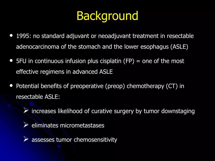
Background • 1995: no standard adjuvant or neoadjuvant treatment in resectable adenocarcinoma of the stomach and the lower esophagus (ASLE) • 5FU in continuous infusion plus cisplatin (FP) = one of the most effective regimens in advanced ASLE • Potential benefits of preoperative (preop) chemotherapy (CT) in resectable ASLE: • increases likelihood of curative surgery by tumor downstaging • eliminates micrometastases • assesses tumor chemosensitivity
Objectives • To determine whether preop FP improves the outcome of resectable ASLE • Primary endpoint: overall survival (OS) • Secondary endpoints: • disease-free survival (DFS) • R0 resection rate • safety
Eligibility Criteria • Histologically-proven adenocarcinoma of the lower third of the esophagus or esogastric junction or stomach • Suitable for curative resection • Age: 18-75 years • WHO performance status (PS): 0 or 1 • Stage II or greater • Non-metastatic disease • Adequate renal and hematological functions • Informed consent
Staging • Endoscopy • Barium meal study • Abdominal and thoracic CT scans • Endoscopic ultrasonography : optional
Study Design Randomization S CT + S FP (*) x 2/3 every 28 days Within 4 weeks 4 - 6 weeks Resection Resection 4 – 6 weeks FP x 3/4 or no treatment Follow-up (*) FP = 5FU: 800 mg/m² CI x 5 days - CDDP: 100 mg/m² at d1 or d2, 1-hr infusion
Postoperative CT: protocol guidelines • Curative surgery • Tumor response* or stabilization after preop CT • pT3 and/or pN+ tumor • No grade 3-4 toxicity under preop CT * based on symptoms (dysphagia, weight loss), endoscopy, CT-scan and +/- endoscopic ultrasonography
Statistical methods • Central randomization stratified on center, PS and tumor site • Sample size: 250patients (178 deaths) to detect an increase from 20% to 35% in 5-yr OS (two-sided logrank test, = 5 %, = 20%) • DFS calculated with a 6 months landmark time to take into account the difference in timing of surgery between the 2 groups • Intent-to-treat analysis • Kaplan-Meier survival curves and two-sided logank-rank test
Recruitment • Open to accrual: November 1995 • Closed to accrual: December 2003 • 224 patients randomized • Eligibility criteria extended in March 1998 to include adenocarcinoma of the stomach
Patient characteristics S CT + S n = 111 n = 113 Median age (yrs) 63 63 (range) (38-75) (36-75) Sex (%) Male 91 (82) 96 (85) Female 20 (18) 17 (15) WHO PS (%) 0 83 (75) 84 (74) 1 28 (25) 29 (26)
Patient characteristics S CT + S n = 111 n = 113 nb pts (%) Site Esophagus 10 (9) 15 (13) Esogastric junction 74 (67) 70 (62) Stomach 27 (24) 28 (25) > 10% weight loss Yes 16 (15) 21 (19) No 88 (79) 87 (77) ND 7 (6) 5 (4)
S N = 111 Preop CT (2-3 cycles) N = 98 (89%) Surgery N = 109 (96%) Surgery N = 110 (99%) Postop CT (1-4 cycles) N = 54 (51%) Trial profile CT + S N = 113
Preoperative CT: Toxicity Grade 3-4 Nb of pts (%) • Neutropenia 22 (20) • Nausea/Vomiting 10 (9) • Thrombocytopenia 6 (5) • Diarrhea 2 (1) • Neurotoxicity 1 (1) • Nephrotoxicity 1 (1) • Mucositis 4 (4) Toxic death 1 (1) Total 40 (37)
Surgery S CT + S n = 111 n = 113 No surgery 1 4* Surgery (%) 110 (99) 109 (96) Median time to surgery (days) 13 78 Postoperative mortality (%) 5 (4) 5 (5) Postoperative morbidity (%) 21 (19) 28 (26) * toxic death (n = 1), disease progression (n = 3)
Type of surgery S CT + S n = 110 n = 109 nb pts (%) No resection 10 (9) 7 (6) Transthoracic esophagectomy 47 (43) 46 (42) Transhiatal esophagectomy 9 (8) 10 (9) Extended gastrectomy 7 (6) 9 (8) Total gastrectomy 21 (20) 23 (21) Distal gastrectomy 14 (13) 15 (14) Other 1 (1) -
Surgical and pathological results S CT + S n = 110 n = 109 nb pts (%) Extent of resection No resection 10 (9) 7 (6) R0 81 (74) 95 (87) p = 0.04 R1 6 (5) 4 (4) R2 12 (11) 2 (2) RX 1 (1) 1 (1)
Pathological results S CT + S p n = 85 n = 98 Tumor stage (%) T0 0 (0) 3 (3) T1, T2 27 (32) 38 (39) T3, T458 (68)57 (58) 0.16 Nodal status (%) N- 17 (20) 32 (33) N+ 68 (80) 66 (67) 0.054 Nb nodes removed median 19 19 range (2 - 82) (1 - 49)
Follow-up and patterns of recurrence • Median follow-up: 5.7 years [2.4-10.4] S CT + S Site of recurrence n = 111 n = 113 nb pts (%) Locoregional only 9 (8) 14 (12) Systemic only 42 (38) 34 (30) Both 20 (18) 14 (12) Total 71 (64) 62 (55)
Logrank p value = 0.0033 Hazard Ratio = 0.65 (95% CI 0.48-0.89) ___ S ___ CT + S years 0-0.5 1 2 3 4 5 6 7 111 57 35 28 21 14 11 6 113 77 53 44 34 25 17 14 At risk Disease-free survival 5-year DFS: 21% (14-30%) vs 34% (26-44%)
Overall survival Logrank p value = 0.021 Hazard Ratio = 0.69 (95% CI 0.50-0.95) ___ S ___ CT + S years At risk 5-year OS: 24% (16-33%) vs 38% (28-47%)
Multivariate & subset analyses • Cox model including: age, gender, PS, tumor site, allocated treatment • Prognostic factors (multivariate analysis): • preop CT (p<0.013) • tumor site (stomach, p <0.01) • No variation of treatment effect according to tumor site (test of interaction, p = 0.26)
Summary of results In resectable gastroesophageal adenocarcinoma,preoperative CT with 5FU/cisplatin: significantly increases curative resection rate significantly improves disease-free survival significantly improves overall survival
Conclusion Our results confirm those of the MAGIC trial* in a slightly different patient population with a non anthracyclin-based CT * Cunningham D et al. N Engl J Med 2006;355:11-20
Participating centers (n=25) Amiens Clermont-Ferrand Montpellier Angers Clichy Nancy Bayonne Colmar Nantes Bicêtre Dijon Reims Bichat Eaubonne Rennes Boulogne Laennec Toulouse Bourg en Bresse Lille Vichy Bourgoin-Jallieu Marseille Villejuif Caen