Download
1 / 105
1.05k likes | 1.06k Views
CENTRAL NERVOUS SYSTEM I NFECT I ONS. Dr. Meral SÖNMEZOĞLU Infectous Diseases Department YEDİTEPE UNIVERSITY HOSPITAL. Learning objectives. Classify central nervous system infections Understand the pathophysiology of central nervous system infections

E N D
CENTRAL NERVOUS SYSTEM INFECTIONS Dr. Meral SÖNMEZOĞLU InfectousDiseasesDepartment YEDİTEPE UNIVERSITY HOSPITAL
Learningobjectives • Classify central nervous system infections • Understand the pathophysiology of central nervous system infections • Know the possible etiologic agents at certain ages • Know the symptoms, signs and diagnosis of central nervous system infections • Know treatment modalities of central nervous system infections • Understand prognosis and know complications • Be alert to the clinical presentation of acute central nervous system infections
CNS Infections • Meningitis • Bacterial, viral, fungal, chemical, carcinomatous • Encephalitis • Bacterial, viral • Meningoencephalitis • Abscess • Parenchymal, subdural, epidural
CNS Infections • Signs and symptoms • Fever • Headache • Altered mental status -lethargy to coma • Neck stiffness – meningismus – flex/ext • Increased intracranial pressure – papilledema, nausea/vomiting, abducens palsies, bulging fontanelle in infants
Meningitis • Bacterial • Viral ( aseptic) • TB • Fungal • Chemical • Parasitic • ? Carcinomatous
Classification of Meningitis • Infectious • Bacterial • Viral • Fungal • Non-infectious • Drug-Induced • Neoplastic • Autoimmune
Noninfectious.. Metabolic Mitochondrial (Reye’s, MELAS) NMS (Neuroleptic malignant syndrome) Nutritional deficiency (Wernicke’s) Paraneoplastic PRES or Malignant hypertension Seizures – (non-convulsive status) TBI Toxic Vascular
BacterialMeningitis • Definition • Bacterial meningitis is an inflammatory response to bacterial infection of the pia-arachnoid and CSF of the subarachnoid space • Epidemiology • Incidence is between 3-5 per 100,000 • More than 2,000 deaths annually in the U.S. • Relative frequency of bacterial species varies with age.
Routes of Entry • Hematogenous • Neighboring focus • Anatomic defect • congenital • traumatic • surgical • Intraneural pathways
Bacterial Meningitis • Streptococcus pneumoniae • Hemophilus influenzae • Listeria moncytogenes • Group B streptococcus • Niesseria meningitidis
Meningitis • Epidemiology • Neonates (< 1 Month) • Gm (-) bacilli 50-60% • Grp B Strep 20-40% • Listeria sp. 2-10% • H. influenza 0-3% • S. pneumo 0-5%
Meningitis • Epidemiology • Children (1 month to 15 years) • H. influenzae 40-60% • Declining dramatically in many geographic regions • N. meningitidis 25-40% • S. pneumo 10-20%
Meningitis • Epidemiology • Adults (> 15 years) • S. pneumonia 30-50% • N. meningitidis 10-35% • Major cause in epidemics • Gm (-) Bacilli 1-10% • Elderly • S. aureus 5-15% • H. influenzae 1-3% • >60 include Listeria, E. coli, Pseudomonas
Meningitis • Pathogenesis • Majority of cases are hematogenous in origin • Organisms have virulence factors that allow bypassing of normal defenses • Proteases • Polysaccharidases
Meningitis • Pathology and Pathogenesis • Sequential steps allow the pathogen into the CSF • Nasopharyngeal colonization • Nasopharyngeal epithelial cell invasion • Bloodstream invasion • Bacteremia with intravascular survival • Crossing of the BBB and entry into the CSF • Survival and replication in the subarachnoid space
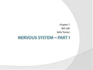
Interrupted arrows indicate where a diffusion of water or solutes can occur between brain capillaries, CSF, and nervous tissue: (a) across the blood-brain barrier; (b) across the epithelium of the choroid plexus; (c) across the ependyma; (d) across the pia-glial membranes at the surface of the brain and spinal cord; (e and f) across the cell membranes of neurons and glial cells. The thick line represents the dura mater and arachnoidea surrounding the system
Pathophysiology of Bacterial Meningitis • Bacterial colonization within the subarachnoid space • Initiation of inflammatory response which leads to: • Endothelial damage • Disruption of the blood-brain barrier • On a larger scale, this results in: • Cerebral edema • Cytotoxic • Vasogenic • Interstitial • Increased ICP
Pathophysiology of Bacterial Meningitis • Pathology • Hallmark • Exudate in the subarachnoid space • Accumulation of exudate in the dependent areas of the brain • Large numbers of PMN’s • Within 2-3 days inflammation in the walls of the small and medium-sized blood vessels • Blockage of normal CSF pathways and blockage of the normal absorption may lead to obstructive hydrocephalus
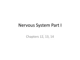
Pathophysiology of Bacterial Meningitis Complications: • Seizures • Hydrocephalus • Infarction • Herniation From van de Beek D Community-acquired bacterial meningitis in adults. 354:1. 44.
ClinicalPresentation of Meningitis • Classicsigns ; • fever, headache, neckstiffness, photophobia, nausea, vomiting, andsigns of cerebraldysfunction (eg, lethargy, confusion, decreased level of consciousness coma). • Thetriad of fever, nuchalrigidity, andchange in mentalstatus is found in onlytwothirds of patients • Atypicalpresentationmay be observed in certaingroups (elderly, diabetic, neutropenic, immunocompromisedhosts..).
ClinicalPresentation of Meningitis • Signs of cerebral dysfunction are common, including confusion, irritability, delirium, and coma. These are usually accompanied by fever and photophobia. • Signs of meningeal irritation are observed in only approximately 50% of patients with bacterial meningitis, and their absence certainly does not rule out meningitis
Meningitis • Clinical Manifestations – Nuchal rigidity • Kernig’s • Pt supine with flexed knee has increased pain with passive extension of the same leg • Brudzinski’s • Supine pt with neck flexed will raise knees to take pressure off of the meninges • Present in 50% of acute bacterial meningitis cases • Cranial Nerve Palsies • IV, VI, VII • Seizures
Amos’s Sign Hips & knees flexed Back arched Neck in extension Trunk supported by arms
Meningitis • Focal neurologic signs may develop as a result of ischemia from vascular inflammation and thrombosis • Papilledema and other signs of increased ICP may be present. • Coma, increased blood pressure with bradycardia, and cranial nerve III palsy may be present. • The presence of papilledema also suggests a possible alternate diagnosis (eg, brain abscess).
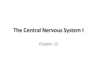
Meningitis Papilledema
Exam in suspected CNS Infection • Mental Status • Cranial nerve and fundiscopic exam • Meningeal Signs • General exam – rashes, lymphadenpathy • Labs – CBCD, BMP, PT/PTT, bHCG, blood cultures, UA C&S • Radiology – CT head - uncontrasted if no focal signs, contrast if mass suspected
Key CSF Features • CSF is not liquid gold – get enough to get your answer • CSF Glucose is 2/3 of serum glucose • Important in diabetic patients • Traumatic LPs – • CSF pro increases by 1 for every 1000 rbcs • Tube #1 and Tube#4 for rbcs when SAH is in the differential not as a routine • Very high CSF Protein levels will make CSF yellow • Send a full tube of CSF for cytology not just a few cc’s
Diagnosis of Meningitis • Diagnosis • Assess for increased ICP • Papilledema • Focal neurologic findings • Defer LP until CT scan or MRI obtained if any of above present • If suspect meningitis and awaiting neuroimaging • Obtain BC’s and start empiric Abx
Diagnosis of Meningitis Obtain CT scan before lumbar puncture in patients with: • Immunucompromised state • History of CNS disease • New onset seizures • Papilledema • Altered level of consciousness • Focal neurologic signs
Diagnosis of Meningitis • Obtain blood cultures and give empiric antibiotics if LP is delayed
LP Increased intracranial pressure is expected – but LP contraindicated if a mass is present or if epidural spinal abscess is suspected Left lateral decubitus position L3-L4 interspace or L4-L5 interspace Think about your studies before the LP
Diagnosing Meningitis • Spinal tap is performed • needle is inserted into an area in the lower back • Identification of the type of bacteria • is important for selection of correct antibiotics.
LP-CSF • Tube # 1 Protein & Glucose • Tube # 2 Gram stain & Culture • Tube # 3 Cell count & differential • Tube # 4 Store ( PCR, viral studies etc)
Diagnosis of Meningitis • Diagnosis • CSF Findings : Opening pressure Appearance Cell count & differential Glucose Protein Gram stain & culture