Download
1 / 7
0 likes | 5 Views
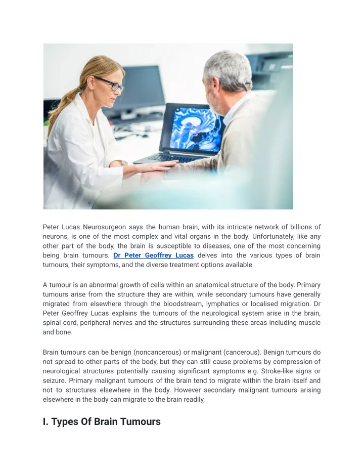
A tumour is an abnormal growth of cells within an anatomical structure of the body. Primary tumours arise from the structure they are within, while secondary tumours have generally migrated from elsewhere through the bloodstream, lymphatics or localised migration. Dr Peter Geoffrey Lucas explains the tumours of the neurological system arise in the brain, spinal cord, peripheral nerves and the structures surrounding these areas including muscle and bone.

E N D
Peter Lucas Neurosurgeon says the human brain, with its intricate network of billions of neurons, is one of the most complex and vital organs in the body. Unfortunately, like any other part of the body, the brain is susceptible to diseases, one of the most concerning being brain tumours. Dr Peter Geoffrey Lucas delves into the various types of brain tumours, their symptoms, and the diverse treatment options available. A tumour is an abnormal growth of cells within an anatomical structure of the body. Primary tumours arise from the structure they are within, while secondary tumours have generally migrated from elsewhere through the bloodstream, lymphatics or localised migration. Dr Peter Geoffrey Lucas explains the tumours of the neurological system arise in the brain, spinal cord, peripheral nerves and the structures surrounding these areas including muscle and bone. Brain tumours can be benign (noncancerous) or malignant (cancerous). Benign tumours do not spread to other parts of the body, but they can still cause problems by compression of neurological structures potentially causing significant symptoms e.g. Stroke-like signs or seizure. Primary malignant tumours of the brain tend to migrate within the brain itself and not to structures elsewhere in the body. However secondary malignant tumours arising elsewhere in the body can migrate to the brain readily, I. Types Of Brain Tumours
Brain tumours are broadly categorized into two types: benign and malignant. Benign Brain Tumours: These are non-cancerous growths that do not invade nearby tissues. They usually have a defined border and are slow growing or may even stop growing for extended periods of time. Common benign brain tumours include meningiomas, pituitary adenomas, and acoustic neuromas. Malignant Brain Tumours: Malignant or cancerous brain tumours can be aggressive, grow rapidly and often invade surrounding brain tissue. They can originate in the brain (primary) or spread from cancer elsewhere in the body (secondary). Examples of primary malignant brain tumours include glioma with the most aggressive known as Glioblastoma Multiforme. Examples of common secondary brain tumours are those arising from lung, breast, skin and colon. II. Symptoms Of Brain Tumors
Slow growing benign tumours may be asymptomatic for some time, however the symptoms of a brain tumour can vary widely, depending on its size, location, and rate of growth, Dr Peter Lucas Neurosurgeon explains. Some common signs and symptoms include: Headaches: Persistent and severe headaches are common early signs of brain tumours. These headaches may worsen over time and are often more intense in the morning or when lying down. Seizures: Brain tumours can disrupt normal brain function, leading to seizures. Seizures may be partial (affecting a specific part of the body) or generalised (involving the entire body). Some seizures may appear like a small stroke that recovers. Nausea and Vomiting: Increased intracranial pressure caused by a tumour can lead to nausea and vomiting, especially in the morning. Changes in Vision: Tumours near the optic nerve can cause vision problems, including blurred vision, double vision, or loss of peripheral vision. Raised intracranial pressure can put generalised pressure on the optic disc (end of the optic nerve) affecting vision. Movement of the eyes can be affected leading to double vision. Motor and Sensory Changes: Weakness or numbness in the limbs, coordination difficulties, and difficulty with balance and walking can occur and be variable through the day. Personality and Cognitive Changes: Brain tumours may affect cognitive functions, leading to changes in memory, concentration, and mood. Personality changes may also be observed. This is more common in lesions affecting the frontal lobes. Speech and Language Problems: Tumours in areas controlling speech and language can result in difficulties in speaking or understanding language. In some cases, the capacity for speech may be lost. III. Diagnosis The diagnosis of a brain tumour typically involves a series of steps, including: General and targeted history: The neurosurgeon will ask a series of questions to ascertain how long an issue has been there, even if previously unrecognised at the time, seeking to gauge the tempo of any clinical change. Further, deficits that may be subtle and unrecognised may become clearer on questioning.
History from other medical practitioners is collated and utilised. Neurological Examination: A neurosurgeon evaluates a patient’s overall neurological health, looking for signs of motor, sensory, or cognitive deficits. Imaging: Brain imaging through techniques like MRI (Magnetic Resonance Imaging) and CT (Computed Tomography) scans is crucial for identifying the location and size of the tumour. In some cases, other imaging such as PET or SPECT imaging may be required. Previous imaging of the region prior to the patient presenting with symptoms, if available, is reviewed. Biopsy: In some cases, a sample of the tumour is obtained through stereotactic guided biopsy in the operating theatre for detailed analysis and to determine its type and grade and to inform a surgical plan. This generally occurs if it is less clear on imaging what type and grade the lesion is or in very extensive non-surgically resectable lesions to confirm diagnosis IV. Treatment Options Treatment options for brain tumours depend on factors such as the type of tumour, its location, the patient’s overall health and the patients’ personal views when fully informed. Some of the common treatment modalities include: Non-Treatment: Particularly in those of advanced age, with extensive high-grade tumour and/or with recurrent high-grade tumour the option to not proceed with any form of treatment needs to be discussed and facilitated as is appropriate. Usually, a neurosurgeon would involve colleagues within palliative care, though also facilitate the patient discussing their care with another neurosurgeon as a second opinion and oncology colleagues such that they are fully informed prior to embarking on this pathway. Surgery: Surgical removal of the tumour is often the first line of treatment. In cases of benign tumours or accessible malignant tumours, complete removal may be possible. This is termed gross total resection. Unlike for instance skin lesions, a “margin” of normal tissue cannot be taken around the tumour due to the deficits this would cause. Relatively recent advancements allow in glioma surgery for medication to be taken orally pre-operatively that causes tumour cells to fluoresce with filtered light under the
microscope differentiating tumour cells from non-tumour cells as an adjunct to surgical removal. Often in higher grade and deeper tumours (within the brain) gross total resection is not possible and debulking for tissue diagnosis, reduction in symptoms and cyto-reduction (less cells) for oncology treatments is the aim. All tumours, benign or malignant, must be considered possible to recur and depending on the type of tumours surveillance imaging and clinical review becomes important in the months and years ahead. Radiation Therapy: Radiation therapy utilizes high-energy beams to target and destroy cancer cells. It is often used after surgery and/or in cases where complete removal is not possible. There are many types of radiation protocols depending on the type of tumour and its location. Advancements in how it is given and at what dose has reduced the side effect profile considerably over the years. Notably, re-irradiating an area should a tumour recur is generally not possible due to the cumulative side effects of the radiation on the non-tumour tissue around the lesion. Further, prior radiation does make any further surgery technically more challenging and increases rates of infection and delayed wound healing. Chemotherapy: Chemotherapy involves the use of drugs to kill or slow the growth of cancer cells. It is often used for malignant brain tumours, either alone or in combination with radiation. Chemotherapy is classically thought of as an IV infusion though many chemotherapeutics are taken orally. It is possible to put chemotherapy wafers in the surgical field once the tumour (glioma tumours) has been removed to the fullest extent safely possible. This therefore commences the chemotherapy delivery locally exactly where it is required from the time of surgery onwards. The wafers control releases the medication over time and then dissolves. Other Therapies: Targeted Therapy: There is extensive research that has been and continues to be conducted regarding all aspects of brain tumours. These reveal novel targets for possible treatment. As these treatments are tested and become more available it is likely we are moving toward unique treatments based on the individual parameters of an individual’s tumour. These may target gene deletions for instance that arise when tumours form.
Immunotherapy: Emerging as a promising treatment, immunotherapy stimulates the patient’s immune system to fight cancer cells. This process has been widely studied within Melanoma research. A tumour sample is taken and an in vitro (lab) deactivated (no longer tumour) sample of tumour cell surface markers are administered to the patient eliciting an immune response that then also targets remaining in vivo (within the body) tumour cells with those cell surface markers. This is a promising area for all tumours. Supportive Care: Managing symptoms and side effects is crucial to improve the patient’s quality of life. This may involve medications, physical therapy, and psychological support. Most commonly swelling is the issue that causes symptoms most rapidly with brain tumours. A strong steroidal anti inflammatory can considerably reduce symptoms rapidly leading up to treatments and during treatment. These medications do have side effects and need to be used wisely. Anti-seizure medication can have a value in preventing a seizure from occurring or treating seizures if they have already occurred. Physical therapies can be as simple as bed exercises to maintain conditioning in a debilitated patient all the way through to strength and conditioning to restore past fitness. The diagnosis of a brain tumour is often a very stressful time not just for the patient but friends and family. Counseling, advice to ensure sleep/eating patterns are maintained and facilitating professional and non-professional support (individuals own support network) are very important as a neurosurgeon. Navigating the path through the medical system requires guidance also as this can add further unwanted stressors. Clinical Trials: Participation in clinical trials can provide access to experimental treatments and contribute to the advancement of brain tumour research. There are promising advances that assist the person in the trial or someone subsequently. Care must be taken in “selling false hope” with respect to trials. They are exactly as they are named. The trial is based on robust research and the hypothetical expectation that they make a difference. Obviously not all trial treatments are of value and it is worth noting that in randomised trials usually half of the cohort receives the “standard current treatment” as the control arm to compare the novel treatment to. V. Prognosis And Hope The prognosis for brain tumour patients varies widely, depending on the type and stage of the tumour, as well as the success of treatment. While malignant brain tumours are
associated with a higher risk, advances in medical science offer hope. Researchers continue to explore innovative treatments, and early detection can significantly improve outcomes. Conclusion Brain tumours are a formidable challenge, affecting the lives of countless individuals, their friends and loved ones. Understanding the types, recognizing symptoms, and knowing the available treatment options are essential in the fight against these complex and potentially life-threatening conditions. “As research and medical technology continue to advance, there is hope for improved treatments and outcomes for those affected by brain tumours with each advancement”, Dr Peter Lucas Neurosurgeon concludes.