Download
1 / 47
530 likes | 1.16k Views
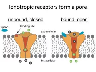
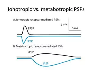
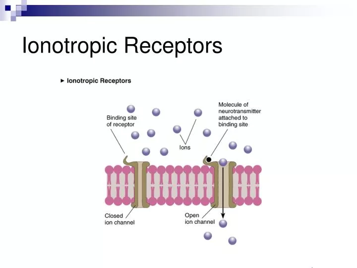
Ionotropic Receptors. Postsynaptic potentials. Depending on the type of ion channel which opens, the postsynaptic cell membrane becomes either depolarized or hyperpolarized.

E N D
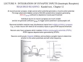
Postsynaptic potentials • Depending on the type of ion channel which opens, the postsynaptic cell membrane becomes either depolarized or hyperpolarized. • Ions will tend to follow the concentration gradient from high to low concentration, and the electrostatic gradient towards the opposite charge.
Excitatory postsynaptic potentials (EPSPs) • Opening of ion channels which leads to depolarization makes an action potential more likely, hence “excitatory PSPs”: EPSPs. • Inside of post-synaptic cell becomes less negative. • Na+ channels (NB remember the action potential) • Ca2+ . (Also activates structural intracellular changes -> learning.) Ca2+ Na+ + outside inside -
Inhibitory postsynaptic potentials (IPSPs) • Opening of ion channels which leads to hyperpolarization makes an action potential less likely, hence “inhibitory PSPs”: IPSPs. • Inside of post-synaptic cell becomes more negative. • K+(NB remember termination of the action potential) • Cl-(if already depolarized) Cl- + outside inside - K+
Neuronal firing: the action potential • The action potential is a rapid depolarization of the membrane. • It starts at the axon hillock and passes quickly along the axon. • The membrane is quickly repolarized to allow subsequent firing.
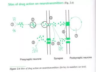
Requirements at the synapse For the synapse to work properly, six basic events need to happen: • Production of the Neurotransmitters • Synaptic vesicles (SV) • Storage of Neurotransmitters • SV • Release of Neurotransmitters • Binding of Neurotransmitters • Lock and key • Generation of a New Action Potential • Removal of Neurotransmitters from the Synapse • reuptake
Overview • Course introduction • Neural Processing: Basic Issues • Neural Communication: Basics • Vision, Motor Control: Models
Motor Control Basics • Reflex Circuits • Usually Brain-stem, spinal cord based • Interneurons control reflex behavior • Central Pattern Generators • Cortical Control
Hierarchical Organization of Motor System • Primary Motor Cortex and Premotor Areas
Primary motor cortex (M1) Hip Trunk Arm Hand Foot Face Tongue Larynx
postsynaptic neuron science-education.nih.gov
Flexor- Crossed Extensor Reflex (Sheridan 1900) Reflex Circuits With Inter-neurons Painful Stimulus
The discovery of mirror neurons in the frontal lobes of monkeys, and their potential relevance to human brain evolution — which I speculate on in this essay — is the single most important "unreported" (or at least, unpublicized) story of the decade. I predict that mirror neurons will do for psychology what DNA did for biology: they will provide a unifying framework and help explain a host of mental abilities that have hitherto remained mysterious and inaccessible to experiments. Ramachandran, Reality Club Lecture 2001
What are mirror neurons? • What is the promise? Why the excitement? • What challenges are faced in fulfilling that promise?
Action observation Action execution Gallese et al. 1996 F5 mirror neurons Shared goal-simulation = Action understanding
Representations in the premotor cortex (Rizzolatti et al). Shift from thinking about movement representations to action representations. Neurons in F4, F5 coding action primitives such as grasping, pinching, pulling
Goal-related neuron in area F5 AGrasping with the mouth BGrasping with the cl. hand CGrasping with the ipsil. hand (Rizzolatti et al. 1988)
90’s: Shift to perceptual responses of F5 neurons Three classes of neurons 1. movement/action neurons Respond only when animal moves 2. “canonical” neurons Respond when object is presented alone 3. mirror neurons Respond when observing action towards object. Same neurons activated during production and perception of an action.
F5 Mirror Neurons A: Effective Action B:Mimicked Action C: Action with tool Gallese et al. Brain 1996
A: Full vision to object B: Hand fades C: Full vision, no object D: Hand fades, no object Umiltà et al. Neuron 2001
Audio-Visual Mirror Neurons Vision+Sound Vision alone Sound alone Movement Kohler et al. Science (2002)
F5 Canonical Neurons Murata et al. J Neurophysiol. 78: 2226-2230, 1997
A New Picture Rizzolatti et al. 1998
The fronto-parietal networks Rizzolatti et al. 1998
F5c-PF Rizzolatti et al. 1998
The F5c-PF circuit Links premotor area F5c and parietal area PF (or 7b). Contains mirror neurons. Mirror neurons discharge when: Subject (a monkey) performs various types of goal-related hand actions and when: Subject observes another individual performing similar kinds of actions
Somatotopy of Action Observation Foot Action Hand Action Mouth Action Buccino et al. Eur J Neurosci 2001
MEG study comparing pianists and non-pianists. Pianists show activation in primary motor cortex when listening to piano. Activation is specific to fingers used to play the notes. Colored region: MEG signal for pianists minus non-pianists.
Physiology of Color Vision Two types of light-sensitive receptors Cones cone-shaped less sensitive operate in high light color vision Rods rod-shaped highly sensitive operate at night gray-scale vision © Stephen E. Palmer, 2002
How They Fire • No stimuli: • both fire at base rate • Stimuli in center: • ON-center-OFF-surround fires rapidly • OFF-center-ON-surround doesn’t fire • Stimuli in surround: • OFF-center-ON-surround fires rapidly • ON-center-OFF-surround doesn’t fire • Stimuli in both regions: • both fire slowly