Download
1 / 29

E N D
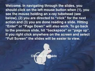
Dr. Norman Ackerman served the University of Florida, College of Veterinary Medicine with distinction as Professor of Radiology from 1979 to 1994. A concerned teacher of veterinary students and residents of all disciplines, Dr. Ackerman also reached the veterinary scientific community through his writing. His numerous clinically pertinent publications are still today a vital part of the veterinary literature; therefore, it is appropriate this site perpetuates Dr Ackerman’s dedication to teaching. This site is presented in recognition of Dr. Norman Ackerman and his contributions to the field of veterinary diagnostic imaging. Sponsorship of the display supports the Dr. Norman Ackerman Memorial Fund, dedicated to the teaching of diagnostic imaging residents at the University of Florida College of Veterinary Medicine. Next slide
Norman Ackerman Memorial Radiography Case Challenge • 10 years old MI German Shepherd dog Next Slide
Signalment • 2 day history of lethargy and inappetence; • Mildly elevated ALT. • You ordered thoracic radiographs Next Slide
Previous Slide Next Slide
Next Slide Previous Slide
Previous Slide Next Slide
Based on your assessment of the radiographs, the thoracic body wall (including the osseous structures and portion of the cranial abdomen included in the collimation) is: • Abnormal • Normal
Correct! There areS4-5 subluxation (white circle) and multifocal sternebral degenerative changes. Next Slide
Sorry! There are S4-5 subluxation (white circle) and multifocal sternebral degenerative changes. Click here to proceed to the next question
Based on your assessment of the radiographs, the pleural space is: • Normal • Abnormal
Correct! There are no abnormalities associated with the pleural space. Next Slide
Sorry! The pleural space is normal Click here to proceed to the next question
Based on your evaluation, the cardiac silhouette is: • Normal • Abnormal
Sorry…almost The cardiac silhouette is within normal limitsfor size and shape, however it is mildly deviated to the right Click here to continue
Correct! There are no abnormalities of size and shape associated with the cardiac silhouette, however it is mildly deviated to the right Next slide
Based on your assessment, the remainder of the mediastinal structures (excluding the cardiac silhouette) are: • Normal • Abnormal
Sorry! • There is an abnormality associated with the mediastinum. Try again!
Correct! • In the cranioventralmediastinal reflection, just dorsal to the second sternebra, there is a 7.5 cm round, well-defined soft tissue opaque mass which causes widening of the cranial mediastinum. Continue
Based on your assessment of the radiographs, the lungs, including the pulmonary vessels, are: • Normal • Abnormal
Sorry! • There is an abnormality associated with the lungs. Try again!
Correct! There is mild diffuse reticular to linear increase in soft tissue opacity throughout the lung lobes, that partially hampers the evaluation of the pulmonary vasculature. Next slide
Conclusion Your findings now include: • Cranioventralmediastinalmass • Mild diffuse unstructured interstitial pulmonary pattern – more likely due to senile pulmonary interstitial fibrosis • Possible S4-5 subluxation and multifocal sternebral degenerative changes. • What are your differential diagnosis for the cranioventralmediastinal mass? click next.
Conclusion • Sternal lymphadenopathy; • Atypically located thymoma, thymiclymphoma, histiocyticsarcoma, or ectopic thyroid tissue cannot be excluded. • Then, Abdominal US + US-guided FNA of the cranial mediastinal mass were performed. click next.
US images click next.
US report • Within the gallbladder, there is a moderate amount of gravity dependent echogenic material; • There is a heterogeneous mildly enlarged mesenteric lymph node measuring 2.5 x 1.1 x 0.7 cm; • Within the cranial mediastinum, there is a hypoechoic mass. This mass has no blood flow as seen with color flow Doppler. click next.
US impressions • Cranial mediastinal mass; consider neoplasia such as thymoma, thymic lymphoma, histiocytic sarcoma, or ectopic thyroid tissue; • Mesenteric lymphadenopathy; consider reactive or neoplastic etiologies; • Gallbladder sludge. The possibility of cholecystitis cannot be ruled out. click next.
FNA results • Thymoma click next.
Take Home Message 1 • In this case, the shape, opacity, and location of the cranial mediastinal mass is likely to be related to sternal lymphadenomegaly; • In a recent study, neoplastic disease was observed in association with sternal lymph node enlargement in approximately 79% of dogs and 69% in cats (Smith & O’Brien, 2012 - JAAHA); • The sternal lymph nodes drain approximately four-fifths of the peritoneal space, as well as portions of the sternal, costal and lumbar areas (Smith & O’Brien, 2012 - JAAHA). That is why is so important to perform abdominal US in these cases, in order to find the underline cause of the possible sternal lymph node enlargement!! click next.
Take Home Message 2 • Due to its rounded, homogeneous and hypoechoic appearance on the US examination, thymoma, thymic lymphoma, and others cranial mediastinal cyst-like lesions were placed higher on the differential diagnostic list; • Usually, thymicneoplasias are diagnosed in mid age to senile dogs (Faiscaet al., 2011 - JSAP), and, in one study, German Shepherd dogs and Labrador Retrievers were over-represented (Day, 1997 - JSAP). Return to the webpage Return to the beginning of the case