Download
1 / 35
350 likes | 496 Views
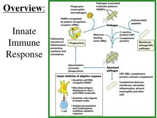
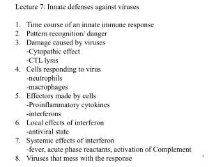
Innate Immune Response. Chapter 15. What am I?. Overview of Innate Defenses. First line of defense are barriers that shield interior of body from external surroundings Anatomical barriers include skin and mucous membranes Provide physical separation

E N D
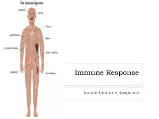
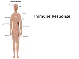
Innate Immune Response Chapter 15
Overview of Innate Defenses • First line of defense are barriers that shield interior of body from external surroundings • Anatomical barriers include skin and mucous membranes • Provide physical separation • Membranes bathed in antimicrobial secretions • Antimicrobial substances and Normal Flora • pH changes
First Line of Defense • Skin • Provides the most difficult barrier to penetrate • Composed of two main layers • Dermis • Contains tightly woven fibrous connective tissues • Makes extremely tough • Epidermis • Composed of many layers of epithelial cells • As cells reach surface, they become increasingly flat • Outermost sheets of cells embedded with keratin • Makes skin water-repellent • Outer layers slough off, taking microbes with it
First Line of Defense • Mucous membranes • Constantly bathed with mucus • Help wash surfaces • Some mucous membranes have mechanisms (cilia)to propel microorganisms and viruses to areas where they can be eliminated
First Line of Defense • Antimicrobial substances • Both skin and mucous membranes are protected by variety of antimicrobial substances including • Lysozyme • Enzymes that degrade peptioglycan • Found in tears, saliva, blood and phagocytes • Peroxidase • Found in saliva, body tissues and phagocytes • Breaks down hydrogen peroxide to produce reactive oxygen • Lactoferrin • Sequesters iron from microorganisms • Iron essential for microbial growth • Found in saliva, some phagocytes, blood and tissue fluids • Defensins • Antimicrobial peptides inserted into microbial membrane • Found on mucous membranes and in phagocytes
First Line of Defense • Normal Microbiota (Flora) • Defined as microorganisms found growing on body surfaces of healthy individuals • Not technically part of immune system • However, provides significant protection • Protects through competitive exclusion • Covers binding sites • Pathogens can’t bind • Competes for nutrients • Nutrients unavailable for pathogens
Cells of the Immune System • Always found in normal blood • Numbers increase during infection • Some cells play dual roles in both innate and adaptive immunity • Blood cell formation called hematopoiesis • Blood cells including immune cells originate from hematopoietic stem cells in bone marrow • Blood cells stimulated to differentiate by colony-stimulating factor
Cells of the Immune System • General categories of blood cells • Red blood cells (RBC) • a.k.a erythrocytes • Carry oxygen in blood • Platelets • Fragments of megakaryocytes • Important component in blood clotting • White blood cells (WBC) • a.k.a leukocytes • Important in host defenses • Divided into four categories • Granulocytes - Mononuclear phagocytes • Dendritic cells - Lymphocytes
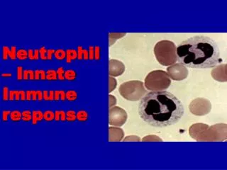
Granulocytes • Contain cytoplasmic graduals • Divided into three types • Neutrophils • Basophils • Eosinophils
Cells of the Immune System • Granulocytes • Contain cytoplasmic graduals • Neutrophils • Most abundant and important in innate response • Granules contain chemicals which kill microbes • Sometimes called polymorphonuclearneutrophilic leukocytes (PMNs)
Cells of the Immune System • Basophils • Granules contain histamine and other chemicals which increase capillary permeability. • Similar to mast cells • Involved in allergic reaction • Eosinophils • Important in expelling parasitic worms • Active in allergic reactions • Granules contain histamase and antimicrobial chemicals
Cells of the Immune System • Mononulcear phagocytes • Constitute collection of phagocytic cells called mononuclear phagocyte system • Include monocytes • Circulate in blood • Macrophages differentiate from monocytes • Present in most tissues • Abundant in liver, spleen, lymph nodes, lungs and peritoneal cavity • Dendritic cells • Branched cells involved in adaptive immunity • Function as scout in tissues • Engulf material in tissue and bring it to cells of adaptive immunity
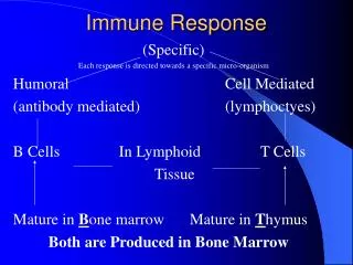
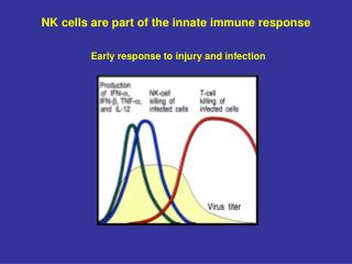
Cells of the Immune System • Lymphocytes • Involved in adaptive immunity • Two major groups • B lymphocytes • B cells-mature in bone marrow • T lymphocytes • T cells-mature in the thymus • Another type • Natural killer • Lacks specificity of B and T cells
Cell Communication • Surface receptors • Membrane proteins to which signal molecules bind • Receptors specific to molecule to which it bonds • Binding molecules called ligands • When ligand binds, receptor becomes modified and sends signal to cell • Cell responds by initiating some action
Cell Communication • Cytokines • Cytokines bind to surface receptors and regulate cell function • Numerous cytokine classes • Chemokines – important in chemotaxis • Colony stimulating factors – Important in multiplication and differentiation of leukocytes • Interferons – important in control of viral infections • Interleukins – produced by leukocytes • Tumor necrosis factor – kill tumor cells
Cell Communication • Adhesion molecules • Allow cells to adhere to each other • Responsible for the recruitment of phagocytes to area of injury • Epithelia cells lining blood vessels produce adhesion molecules that catch phagocytes as they pass by • Cause phagocytes to slow and leak out of vessels to area of injury
Sensor Systems • Systems within blood detect signs of tissue damage or microbial invasion • Respond to patterns associated with danger by • Directly destroying invading microbe • Recruiting other host defenses
Sensor Systems • Toll-like receptors (TLR) and NOD proteins • Pattern recognition receptors • TLR allow cells to “see” molecules signifying presence of microbes outside the cell • TLR found in variety of cell types • Recognize distinct “danger” compounds • Signal is transmitted • Results in change of gene expression of cell • NOD proteins do same for inside cell
Sensor Systems • Complement system • Series of proteins circulating in blood and fluids • Circulate in inactive form • Augment activities of adaptive immune response • Stimulation of inactive proteins initiates cascade of reactions • Results in rapid activation of components • Three pathways of activation • Alternative pathway • Lectin pathway • Classical pathway
Sensor Systems • Classical pathway • Activation requires antibodies • Antibodies interact complement C1 • Activates protein • Leads to activation of all complex proteins • Alternative pathway • Quickly and easily initiated • Relies on binding of complement protein C3b to cell surface • Initiates activation of other compliment proteins • Allows formation of complement complex • C3b always circulating in blood
Sensor Systems • Lectin pathway • Activation requires mannan-binding lectins (MBL) • Pattern recognition molecules • Detect mannan • Polymer of mannose • Found in microbial cells • MBL attaches to surface • Activates complement proteins
Sensor Systems • Lysis of foreign cells • Complexes of C5b, C6, C7, C8 and multiple C9 spontaneously assemble • Forms donut-shaped structure called membrane attack complex (MAC) • Creates pores in membrane • Most effective on Gram-negative cells • Little effect on Gram-positive cells
Sensor Systems • Long Double-Stranded RNA (dsRNA) • Induction of alpha and beta interferons • Cells express iAVPs • Leads to apoptosis
Phagocytosis • Process of phagocytosis • Chemotaxis • Cells recruited to infection • Recognition/attachment • Use receptors to bind invading microbes • Engulfment • Phagocyte engulfs invader-forming phagosome • Phagosome lysosome fusion • Phagosome binds lysosome, forming phagolysosome • Destruction and digestion • Organism killed due to lack of oxygen and decreased pH • Exocytosis • Phagocyte expels material to external environment
Phagocytosis • Role of Neutrophiles • First responders • Granules contain antimicrobial chemicals • NETs-neutrophile extracellular traps • Contain DNA and anti microbial chemicals • Trap bacteria and destroy them with chemicals • Short lived but lots in reserve
Inflammation • Inflammation occurs in response to tissue damage • Four cardinal signs • Heat • Pain • Redness • Swelling • Loss of function • Fifth sign that can also be present
Inflammation • Factors that initiate inflammatory response • Microbial products trigger toll-like receptors of macrophages • Causes release of pro-inflammatory cytokines • Microbial cell surface can trigger complement • Tissue damage results in enzymatic cascade • Cascades initiate inflammation
Inflammation • The inflammatory process • Initiation leads to a cascade of events • Results in dilation of blood vessels, leakage of fluid from vessels and migration of leukocytes and phagocytes • Leakage of phagocytes from blood vessels called diapedesis • Certain pro-inflammatory mediators cause the diameter of blood vessels to increase • Results in increased blood flow • Increased blood flow responsible for cardinal signs of inflammation
Inflammation • Outcomes of inflammation • Intent is to limit damage and restore function • Inflammation itself can cause considerable damage • Release of toxic products and enzymes from phagocytic cells is responsible for tissue damage • If inflammation is limited to area of injury, damage is usually nominal • If inflammation results in delicate systems, consequences are more severe • Inflammation around brain and spinal cord can lead to meningitis
Inflammation • Apoptosis • Programmed cell death • Destroys cell without eliciting inflammatory response • During apoptosis, cells undergo changes to signal macrophages • Cells are engulfed without triggering inflammatory cascade
Fever • One of the strongest indicators of infection • Especially of bacterial infection • Important host defense mechanism • Temperature regulation center of body responds to fever-inducing substances called pyrogens • Fever-inducing cytokines termed endogenous pyrogens • Microbial products termed exogenous pyrogens • Resulting fever inhibits growth of pathogens by • Elevating temperature above maximum growth temperature • Activating and speeding up other body defenses