Download
1 / 59
630 likes | 809 Views
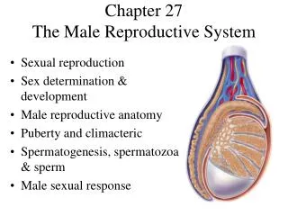
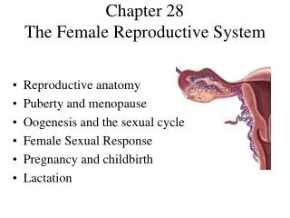
The Reproductive System and Development Chapter 25 – Lecture Notes. to accompany Anatomy and Physiology: From Science to Life textbook by Gail Jenkins, Christopher Kemnitz, Gerard Tortora. Chapter Overview. 25.1 Sperm Production 25.2 Male Reproductive System

E N D
The Reproductive System and DevelopmentChapter 25 – Lecture Notes to accompany Anatomy and Physiology: From Science to Life textbook by Gail Jenkins, Christopher Kemnitz, Gerard Tortora
Chapter Overview 25.1 Sperm Production 25.2 Male Reproductive System 25.3 Female Reproductive System 25.4 Vagina and Mammary Glands 25.5 Ovarian and Uterine Cycle 25.6 Implantation of Blastocyst 25.7 Embryonic to Fetal Period
Chapter Overview 25.8 Maternal Changes During Pregnancy 25.9 Labor 25.10 Milk Production and Ejection
Essential Terms gamete • germ cells with haploid number of chromosomes fertilization • occurs when sperm unites with secondary oocyte pregnancy • sequence of events resulting in birth of child gonads • male testes and female ovaries which secrete hormones and produce gametes
Introduction Sexual Reproduction • Males and females differ anatomically to produce gametes and support a fetus • Fertilization is the result of male and female gametes joining • Pregnancy begins with fertilization and usually results in birth of a child • Gonads produce gametes and hormones
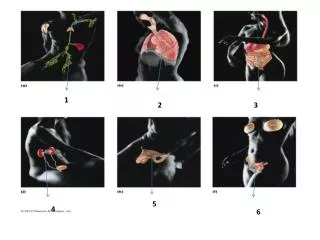
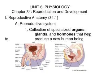
Male Reproductive System • Testes – System of Ducts • Epididymis, ductus deferens, ejaculatory ducts, and urethra • Accessory glands • Seminal vesicles, prostate, and bulbourethral glands • Supporting Structures • Scrotum and penis
Scrotum • Scrotal location and muscle contraction regulate temperature of testes • Normal sperm production occurs 2-3°C lower than core body temperature • Response to cool temperatures • Cremaster muscles contract to pull testes close to body • Dartos muscles contract to tighten scrotum • Response to warm temperatures • Reverse of above actions
Testes • One testes in each sac of scrotum • Descend during seventh month of fetal development • Tunica albuginea is located deep to tunica vaginalis and forms 200-300 lobules • Lobules contain seminiferous tubules • Spermatogenesis carried out by seminiferous tubules of testes
Testes • Spermatogenic cells • Begin sperm production at puberty • Sertoli or sustentacular cells • Junctions form blood-testis barrier • Nourish spermatogenic cells • Carry out phagocytosis • Control spermatogenic movement • Produce fluid for transport • Secrete hormone inhibin • Leydig (interstitial) cells • Secrete testosterone
Spermatogenesis • Begins with spermatogonia and diploid number of chromosomes • Some pass through blood-testis barrier • Primary spermatocytes also diploid
Spermatogenesis • Meiosis I • Crossing-over during metaphase I • Secondary spermatocytes result • Each with haploid number • Meiosis II • Results in four spermatids • Each with haploid number
Spermatogenesis • Cytoplasmic bridges link the four daughter cells • Spermiogenesis • Transformation of spherical spermatids into elongated sperm • Formation of arcosome • Flagellum develop • Mitochondria multiply
Sperm • 300 million per day produced • Survive 48 hours in female reproductive tract • Sperm Parts • Head • Nucleus • Arcosome • Tail • Neck • Middle piece • Principal piece • End piece
Hormonal Control of Testes • Negative feedback loops control testosterone release and spermatogenesis • Gonadotropin-releasing Hormone (GnRH) • Increased production at puberty • Stimulates secretion of LH and FSH • Luteinizing hormone (LH) • Stimulates Leydig cells • Follicle-stimulating hormone (FSH) • Stimulates Sertoli cells to secrete ABP
Hormonal Control of Testes • Testosterone • Principal androgen • Synthesized from cholesterol • Suppresses LH and GnRH secretion • Converted to DHT in prostate and seminal vesicles • Dihydrotestosterone (DHT) • Stimulates development of external genitals • Androgen-binding Protein (ABP) • Binds to and keeps testosterone levels high • Inhibin • Inhibits FSH secretion
Effects of Testosterone and Dihydrotestosterone • Prenatal development • Development of male sexual characteristics • Development of sexual function • Stimulation of anabolism
Reproductive System Ducts in Males • Ducts of testis • Seminiferous tubules • Straight tubules • Rete testis • Efferent ducts • Ductus epididymis • Epididymis • Site of sperm maturation and storage • Consists of ductus epididymis • Head – superior portion formed by efferent ducts of testis • Body • Tail – continues as ductus (vas) deferens
Ductus Deferens • Terminal portion is the ampulla • Conveys sperm from epididymis toward urethra • Storage and reabsorption of sperm
Spermatic Cord • Supporting structure ascends out of scrotum • Comprised of several structures • Ductus deferens • Testicular artery • Pampiniform plexus • Autonomic nerves • Lymphatic vessels • Cremaster muscles
Ejaculatory Ducts • Formed by duct of seminal vesicle and ampulla of ductus deferens • Eject sperm and seminal vesicle secretions from urethra to exterior
Urethra • Shared by reproductive and urinary systems • 3 subdivisions • Prostatic urethra • Membranous urethra • Spongy (penile) urethra
Accessory Sex Glands • Produce fluids to protect semen and facilitate their movement • Seminal vesicles • Prostate • Bulbourethral or Cowper’s glands
Seminal Vesicles • Alkaline fluid • Fructose • Prostaglandins • Clotting proteins • Approximately 60% of semen volume
Prostate Gland • Citric acid • Proteolytic enzymes – prostate-specific antigen (PSA) • Pepsinogen • Lysozyme • Amylase • Hyaluronidse • Milky, acidic fluid (pH ~ 6.5) • Approximately 25% semen volume
Bulbourethral / Cowper’s Glands • Pea sized glands active during sexual arousal • Alkaline fluid • Secrete lubricating mucus
Semen • Combination of sperm and seminal fluid • 2.5 – 5 mL per ejaculation • 50 – 150 million sperm/ml • Alkaline pH ~ 7.2 – 7.7 • Seminalplasmin – antibiotic
Penis • Contains urethra • Passage for sperm and urine • 3 Parts • Root • Body • Glans penis • Erection maintained by parasympathetic fibers • Ejaculation, a sympathetic reflex, releases semen to exterior