Download
1 / 69
690 likes | 778 Views
Explore the different types of muscles - smooth, cardiac, and skeletal - and their functions in body movement, heat production, and more. Learn about the structure of skeletal muscle fibers and the process of muscle contractions.

E N D
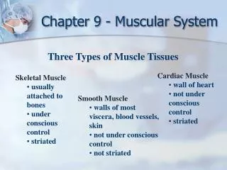
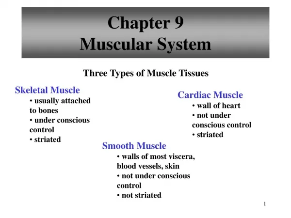
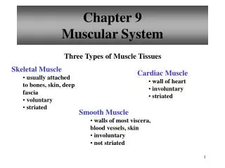
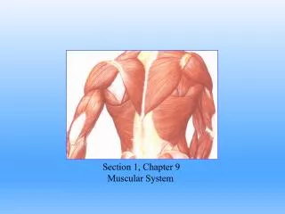
Section 1, Chapter 9 Muscular System
Muscle is derived from Musculus, for “Mouse” Imagine a mouse running beneath the skin. Functions of Muscles: Body movement Maintain posture Produces heat Propel substances through body Heartbeat Types of muscles include: Smooth muscle Cardiac muscle Skeletal muscle
Smooth Muscle • Characteristics of smooth muscles • Involuntary control • Tapered cells with a single, central nucleus • Lack striations
Smooth Muscle There are two types of smooth muscles • Multi-unit Smooth Muscle • unorganized cells that contract as individual cells • Located within the iris of eye and the walls of blood vessels • Visceral (single-unit) Smooth Muscle • Form sheets of muscle • Cells are connected by gap junctions • Muscle fibers contract as a group • Rhythmic contractions • Within walls of most hollow organs (viscera)
Cardiac Muscle • Located only in the heart • Striated cells • Intercalated discs • Muscle fibers branch • Muscle fibers contract • as a unit • Self-exciting and rhythmic
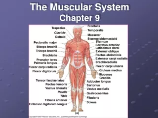
Skeletal Muscle • Usually attached to bone • Voluntary control • Striated (light & dark bands) • Muscle fibers form bundles • Several peripheral nuclei
Coverings of Skeletal Muscle • Fascia • Dense connective tissue surrounding skeletal muscles • Tendons • Dense connective tissue that attaches muscle to bones • Continuation of muscle fascia and bone periosteum • Aponeurosis • Broad sheet of connective tissue attaching muscles to bone, or to other muscles.
Coverings of Skeletal Muscle • Epimysium • Connective tissue that covers the • entire muscle • Lies deep to fascia • Perimysium • Surrounds organized bundles of • muscle fibers, called fascicles • Endomysium • Connective tissue that covers • individual muscle fibers (cells)
Figure 9.3 Scanning electron micrograph of a fascicle surrounded by its perimysium. Muscle fibers within the fascicle are surrounded by endomysium.
Organization of Skeletal Muscle • Fascicle • Organized bundle of muscle fibers • Muscle Fiber • Single muscle cell • Collection of myofibrils • Myofibrils • Collection of myofilaments • Myofilaments • Actin filament • Myosin filament Figure 9.2 Skeletal muscle organization
Skeletal Muscle Fibers • Sarcolemma • Cell membrane of muscle fibers • Sarcoplasm • Cytoplasm of muscle fibers • Sarcoplasmic Reticulum • Modified Endoplasmic Reticulum • Stores large deposits of Calcium sarcolemma
Skeletal Muscle Fibers • (Transverse)T-tubules: • invaginations of sarcolemma, • extending into the sarcoplasm. • Cisternae: • enlarged region of sarcoplasmic • reticulum, adjacent to the t-tubules • Triad • T-tubule + adjacent cisternae Openings into t-tubules
Myofibrils • Myofibrils are bundles of actin and myosin filaments. • Actin – thin filament • Myosin – thick filament • Striations appear from the organization of actin and myosin filaments Figure 9.4 Organization of actin and myosin filaments
Sarcomere • A sarcomere is the functional unit of skeletal muscle • A sarcomere is the area between • adjacent Z-lines. • During a muscle contraction the z-lines move together and the sarcomere shortens.
Striations appear from alternate light and dark banding patterns. Z Line is the attachment site of actin filaments (center of I bands) • I Bands (light band): consists of only • actin filaments • A Bands (dark band) : consists of myosin filaments and the overlapping portion of actin filaments Figure 9.5 thin and thick filaments in a sarcomere.
filaments • Thin filaments • composed of actin proteins • Thin filaments are associated with troponin • and tropomyosin proteins • Thick filaments • composed of myosin proteins • During muscle contraction the heads on myosin filaments bind to actin filaments forming a Cross-bridge
Cross-Bridges When a muscle is at rest, myosin heads are extended in the “cocked” position. During a contraction, myosin heads bind to actin, forming a cross-bridge and the myosin head pivot forward (Power Stroke) and back (Recovery stroke)
Troponin-Tropomyosin Complex The troponin-tropomyosin complex prevents cross-bridge formation when the muscle is at rest. • Tropomyosin • Blocks binding sites on • actin when a muscle is • at rest • Troponin • Ca2+ binds to troponin • during a muscle contraction. • Troponin moves repositions the tropomyosin filaments, so the myosin and actin filaments can interact. End of section 1, chapter 9
Chapter 9, Section 2 Muscle Contractions
Synapse Synapse: Functional (not physical) junction between an axon of a neuron and another cell The two cells are separated by a physical space, called the synaptic cleft. Neurotransmitters are stored within synaptic vesicles of the presynaptic cell and they’re released into the synapse.
Neuromuscular Junction Neuromuscular Junction (NMJ) refers to the synapse between an axon and a muscle fiber. Motor End Plate is a highly folded region of muscle fiber at NMJ that contain abundant mitochondria Figure 9.8a. General NMJ
Motor Unit Motor neurons innervate effectors (muscles or glands) A motor unitincludes a motor neuron and all of the muscle fibers it controls 1 motor unit may control between 1 and 1000 muscle fibers Figure 9.9 two motor units. The muscle fibers of a motor unit are innervated (controlled) by a single motor neuron.
Stimulus for Contraction Acetylcholine (ACh) is the only neurotransmitter that initiates skeletal muscle contraction Sequence of Actions • A nerve impulse (Action Potential) reaches axon terminal • The impulse opens calcium channels at the axon terminal • Calcium diffuse into axon • The calcium triggers the release of ACh from vesicles into synaptic cleft.
Stimulus for Contraction Sequence of Actions…Continued ACh diffuses across synaptic cleft & binds to receptors on motor endplate. ACh opens Na+ channels on muscle Na+ floods into the muscle, initiating a muscle impulse. A muscle impulse (action potential) is propagated across the entire muscle.
Stimulus for a muscle impulse. Corresponds to steps 1-7 in the previous slides.
The muscle impulse causes the release of calcium from the SR. Calcium binds to troponin and tropomyosin is repositioned exposing the actin filaments.
Stimulus for contraction continued… 8. The muscle impulse diffuses across sarcolemma and down the t-tubules into the cisternae of sarcoplasmic reticula. 9. The sarcoplasmic reticula release their calcium supplies into the sarcoplasm. 10. Calcium binds to troponin and the troponin repositions the tropomyosin, so the myosin can bind to actin. 11. Cross-bridge cycling causes the muscle to contract.
Excitation-Contraction Coupling Calcium released from sarcoplasmic reticulum binds to troponin. Troponin moves tropomyosin, exposing actin filaments to myosin cross-bridges. myosin heads bind to actin, forming a cross bridge and cross-bridge cycling causes the muscle to contract. End of section 2, chapter 9
ivyanatomy.com section 3, chapter 9 Sliding Filament Theory of Contraction
The Sliding Filament Model of Muscle Contraction • During a muscle contraction • Thick (myosin) filaments and thin (actin) filaments slide across one another • The filaments do not change lengths • Z-bands move closer together causing the sarcomere to shorten. • I bands appear narrow Figure 9.11a. Individual sarcomeres shorten as thick and thin filaments slide past one another.
Cross Bridge Cycling When a muscle is relaxed, tropmyosincovers the binding sites on actin. A molecule of ADP and Phosphate remains attached to myosin from the previous contraction.
Cross Bridge Cycling • During a contraction, Calcium binds to troponin. • Tropomyosin is repositioned, exposing the myosin binding sites on actin filaments
Cross Bridge Cycling Myosin heads bind to actin filaments. The phosphate is released.
Cross Bridge Cycling Myosin heads spring forward “Power Stroke” pulling the actin filaments. ADP is released from Myosin
Cross Bridge Cycling • Myosin is released from actin. • A new molecule of ATP binds to myosin, causing it to be released from the actin filament. • ATP is not yet broken down, but it is essential to release the cross-bridges.
Cross Bridge Cycling ATP is broken down, providing the energy to “cock” the myosin filaments (recovery stroke). Steps 1-6 are repeated several times.
Figure 9.10. The cross-bridge cycle. The cycle continues as long as ATP is present, and nerve impulses release Acetylcholoine. Watch the You-Tube video “Sliding Filament” to view cross-bridge cycling in action.
Relaxation • When a nerve impulse ceases, two events relax muscle fibers. • Acetylcholinesterase breaks down Ach in the synapse. • Prevents continuous stimulation of a muscle fiber. • Calcium Pumps (Ca2+ATPase) remove Ca2+ from the sarcoplasm and returns it to the SR. • Without calcium, tropomyosin covers the binding sites on actin filaments.
Relaxation * Notice that ATP is required for muscle relaxation!
ivyanatomy.com section 4, chapter 9 Energy Sources for Contraction
Energy Sources for Contraction • ATP provides the energy to power the interaction between actin & myosin filaments. • However, ATP is quickly spent and must be replenished New ATP molecules are synthesized by Hydrolysis of Creatine Phosphate Glycolysis (anaerobic respiration) Aerobic Respiration
Creatine Phosphate Creatine Phosphate can be hydrolyzed into Creatine, releasing energy that is used to make new ATP. The energy from creatine phosphate hydrolysis cannot be used to directly power muscles. Instead, it’s used to produce new ATP.
Creatine Phosphate…continued • When cellular ATP is abundant, creatine phosphate can be replenished by phosphorylating creatine. • Creatine Phosphate provides energy for only about 10 seconds of a high intensity muscle contraction.
Glycolysis Anaerobic respiration (glycolysis) occurs in the cytosol of the cell and does not require oxygen. Glucose molecules are partially broken down producing just 2 ATP for each glucose. If there isn’t sufficient oxygen available, glycolysis produces lactic acid as a byproduct.
Oxygen debt of glycolysis Exercise and strenuous activity depends on anaerobic respiration for ATP supplies. During exercise anaerobic respiration causes lactic acid to accumulate in the cells. After exercise, when oxygen is available the O2 is used to convert lactic acid back to glucose in the liver. Oxygen debt is the amount of oxygen needed by liver cells to convert accumulated lactic acid back to glucose.