Download

1 / 8
80 likes | 88 Views
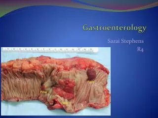
Gastroenterology is the branch of medicine focused on the digestive system and its disorders. Diseases affecting the gastrointestinal tract, which include the organs from mouth into anus, along the alimentary canal, are the focus of this speciality

E N D
vv Clinical Group Archives of Clinical Gastroenterology DOI CC By ISSN: 2455-2283 Luis Angel Medina Andrade1*, Arsenio Torres Delgado2, Luis Fernando Perez Corona2, Oscar Sanchez Moreno3, Daniel Flores Rodríguez4, Oscar Rodrigo Rocha Erazo5, Eduardo Vidrio Duarte6, José Daen Jonathan Cabañas Gomez7, Rene Cano Rodriguez5, Nestor Apaez Araujo7 and Oscar Arturo Nava Carmona8 Case Report Acute Pancreatitis, Actualization and Evidence Based Management Abstract 1General Surgery Department, General Zone Hospital #30, Mexican Social Security Institute, Mexico City, México 2General Surgery Department, Tulancingo General Hospital, Hidalgo, México 3Endoscopy Department, Gea Gonzalez Hospital, México City, México 4General Surgery Department, Veracruz High Specialty Regional Hospital ISSSTE, Veracruz, México 5General Surgery Department, Military School for Sanity Graduated, National Defense Secretary, México City, México 6General Surgery Department, Hospital Angeles Metropolitano, La Salle University, Mexico City, México 7General Surgery Department, Ecatepec General Hospital, México 8General Surgery Department, León General Hospital, México Acute pancreatitis (AP) is a systemic immunoinfl ammatory response to auto-digestion of the pancreas and peri-pancreatic organs. It is a frequent gastrointestinal disease with an important morbi- mortality, reaching 30% in severe cases. Different studies and reviews by international groups have developed multiple classifi cation systems to assess the severity and address the correct management along time, identifying the better molecular markers, clinical outcome determinants and reaching conservative management as the angular piece in AP. In this review we present a compilation of the latest studies and international consensus about AP physiology, etiology, risk factors, diagnosis, severity assessment, imaging and treatment. Pathophysiology There are plenty of theories about the pathophysiology of AP, most of them conclude that distal ductal obstruction, irrespective of the mechanism, leads to upstream blockage of pancreatic secretion, which in turn prevents exocytosis of zymogen granules (containing digestive enzymes) from acinar cells. Consequently, the zymogen granules merge with intracellular lysosomes to form condensing or autophagic vacuoles, containing an admixture of digestive and lysosomal enzymes [3]. Dates: Received: 03 December, 2016; Accepted: 12 January, 2017; Published: 19 January, 2017 *Corresponding author: Luis Angel Medina Andrade, MD, General Surgery Department, General Zone Hos- pital #30, Mexican Social Security Institute, Mexico City, México, E-mail: The activation of this enzymes, normally inactive into the pancreas, produces a proinfl ammatory signals cascade along the gland, with posterior release in the circulatory system and the consequently systemic infl ammatory response syndrome (SIRS). The IL-1, IL-2, IL-6 and IL-8 release favors monocytes and macrophages quimiotaxis and signal amplifi cation with TNF- release by macrophages, with fi nal permeability increase in different systems like vascular, gastrointestinal and the consequent bacterial translocation from gut lumen to the circulation [4]. Keywords: Pancreatitis; Severe pancreatitis; Chole- cystitis complications; Nutritional management https://www.peertechz.com Introduction Acute pancreatitis (AP) is a systemic immunoinfl ammatory response to auto-digestion of the pancreas and peri-pancreatic organs. AP is a common and life threatening disease. Annual incidence worldwide is 4.9–73 cases per 100000 people; affecting young adults, with a male: female ratio of 2.5:1. The mortality rate for pancreatitis is between 1.5% and 4.2% in large epidemiological studies, but varies according to pancreatitis severity, reaching 30% in those with infected pancreatic necrosis [1,2]. Another theory propose bile acids as the pathogenic factor in biliary pancreatitis, when it is taken up by acinar cells from the bile acid transporters in apical and basolateral plasma membrane, leading to intracellular calcium increasing and the consequent increase in transcription of some proinfl ammatory mediators [5]. In the case of alcoholic pancreatitis, it increases digestive and lysosomal enzymes content in acinar cells, destabilizing 001 Citation: Medina Andrade LA, Torres Delgado A, Perez Corona LF, Moreno OS, Rodríguez DF, et al. (2017) Acute Pancreatitis, Actualization and Evidence Based Management. Arch Clin Gastroenterol 3(1): 001-008. DOI: http://doi.org/10.17352/2455-2283.000028
Medina Andrade et al. (2017) the organelles containing enzymes, increasing the probability of contact between lysosomal and digestive enzymes with the consequent high risk of intracellular activation of digestive enzymes [5,13]. Risk factors Multiple risk factors are associated with the development and severity of AP. Diabetes mellitus II have been documented to increase the risk of AP in 1.86-2.89 times [5,14,15,21,]. Once the initial damage is established the progression and outcomes would depend on the medical management during the fi rst 24 hours [6]. In case of limitation of the pancreatitis trigger and the correct initial management with aggressive fl uid therapy, the pancreatic injury and the cytokine release could be limited, with the consequent decrease in SIRS and better outcomes. If it is not accomplished, acinar cells would develop ischemia and necrosis secondary to hipoperfusion [7]. Persistent cytokines release by necrotic tissue would increase vascular permeability favoring pulmonary effusion and respiratory distress, hypovolemia, hypotension, acute renal failure, intestinal edema and intra-abdominal hypertension [8,9]. There is a signifi cative association between body mass index and development of biliary AP. Although there is a high prevalence of metabolic syndrome it has been demonstrated that waist circumference, body mass index, age or sex were not related with pancreatitis severity [22]. In the other hand obesity is a chronic low-grade infl ammatory state characterized by high circulating levels of proinfl ammatory cytokines. Alternatively, obesity may intensify the immune response, which is able to exacerbate pancreatic injury and is related with a poor prognosis [13,22,23]. Current and former smoking are associated with increased risk for AP. Several experimental studies on rat models have investigated the effect of smoking showing increased infl ammatory activity, focal infl ammation, and decreased number of acinar structures and up-regulation of genes expressing digestive enzymes [24-26]. It has been demonstrated in studies by Sadr-Azodi, Tolstrup and Lindkvist et al., that smoking increase the relative risk of non-gallstone related acute pancreatitis 2.29, and was confi rmed that duration is more important that smoking intensity5. Interstitial edema, absent peristalsis and increased gut permeability have been associated with bacterial translocation and sepsis. Intra-abdominal abdominal compartmental syndrome (ACS) can be developed in almost 35% of cases of severe pancreatitis, secondary to intestinal edema, ascites and retroperitoneal liquid collections, sometimes precipitated by an aggressive fl uid therapy. In fact, actually IAH and ACS are considered a severity parameter [9- 13]. hypertension (IAH) and Multiple genetic factors are being studied to elucidate the pathophysiology of AP because some patients with a seemingly mild pancreatic injury (e.g., during endoscopic retrograde cholangiopancreatography [ERCP] without pancreatic duct injection) develop severe AP, whereas other subjects with extensive injury have a relatively mild course. For example, the angiotensin-converting enzyme I (rather than D) allele was signifi cantly associated with alcohol-related AP (p = 0.03). The renin rs5707 G (rather than A) allele was associated with AP (p = 0.002), infected necrosis (p = 0.025) and mortality (p = 0.046)) [27]. In preliminary studies, the authors found that the MCP-1–2518 A/G single nucleotide polymorphism predicted that the physiological response to pancreatitis would be severe and was associated with death [28]. Other mutations in genes like SPINK1, a gene that encodes for a pancreatic trypsin inhibitor, are associated with acute pancreatitis resulting from an impaired ability to counteract the effects of activated trypsin within pancreatic acinar cells. Etiology There are plenty of AP causes (Table 1). Alcohol and gallstones are responsible of 80% of cases. Incidence of idiopathic pancreatitis is increasing, maybe related with risk factors like obesity and metabolic syndrome, and the 57% of this idiopathic cases have been demonstrated with microlithiasis as cause after endoscopic ultrasound or magnetic resonance cholangyopancreatography [14-16]. People with gallstone disease will develop pancreatitis in 5%, and 25% of this will turn in a severe one [17]. Without defi nitive treatment in this cases recurrence is as high as 40% [18]. Choledocholithyasis is uncommon (20%–30%) following a mild attack of ABP [19]. Gallstones <5 mm in diameter are more likely to cause pancreatitis than larger stones. The presentation of acute pancreatitis in only 2-3% of alcoholic people suggest a genetic factor implicated [18]. In this etiology consumption of >100 g of alcohol daily and low intake of fat are signifi cant risk factors [17]. Diagnosis and etiology assessment Table 1: AP causes. The diagnosis of acute pancreatitis is based on the fulfi llment of 2 of the following criteria [29]: Frequent Less Frequent Gallstones Alcoholism Hypertriglyceridemia Post–endoscopic retrograde cholangiopancreatography Idiopathic ( Microlitiasis 57%) Drug induced: Azathioprine, 6-Mercaptopurine, Trimethoprim- sulfamethoxazole, Pentamidine, 2,3 Dideoxyinosine, Asparginase, Methyl- dopa. Autoimmune Genetic Abdominal trauma Postoperative Ischemia Infections Hypercalcemia and hyperparathyroidism Posterior penetrating ulcer Scorpion venom Pancreas divisum -Clinical upper abdominal pain -Serum amylase or lipase >3x upper limit of normal -Computed Imaging (MRI), or ultrasonography diagnosis. Tomography (CT), Magnetic Resonance Once the diagnosis is established the etiology must be elucidated (Table 1), to select the correct management for better 002 Citation: Medina Andrade LA, Torres Delgado A, Perez Corona LF, Moreno OS, Rodríguez DF, et al. (2017) Acute Pancreatitis, Actualization and Evidence Based Management. Arch Clin Gastroenterol 3(1): 001-008. DOI: http://doi.org/10.17352/2455-2283.000028
Medina Andrade et al. (2017) outcomes. The main etiology is gallstones in 40% of cases. Abdominal USG is the preferred imaging study for abdominal pain associated with jaundice and for exclusion of gallstones as the cause of acute pancreatitis. Pancreas use to be visualized inadequately in 30% of cases, with about 50% of sensitivity for the detection of choledocholithyasis. Gallstones <5 mm in diameter are more likely to cause pancreatitis. An ALT> 150UI/L had a positive predictive value of 95% in diagnosing acute gallstone pancreatitis [30-33]. pancreatic necrotic collections and walled-off necrosis (Table 4), while systemic complications can be related to exacerbations of underlying co-morbidities exacerbated by acute pancreatitis. Mild acute pancreatitis is characterized by the absence of organ failure and the absence of local or systemic complications. Moderately severe acute pancreatitis is characterized by the presence of transient organ failure, local or systemic complications in the absence of persistent organ failure [37]. One would expect the presence of a local complication by persistence of abdominal pain, secondary increases in serum amylase/lipase activity, organ failure, fever/chills, and so forth. Such symptoms usually prompt a cross-sectional imaging procedure to search for these complications [38]. Alcohol consumption is the responsible for about 30% of pancreatitis cases. Clinical history can elucidate the origin, like drinking more than eight alcoholic drinks/day (>100g/d) for more than 5 years. It is present in males predominantly with a male: female ratio of 2.5:1, most of them young adults [34]. Table 2: Classifi cation System for severity of Acute Pancreatitis. Proposed mechanisms of alcohol damage include sphincter of Oddi spasm, precipitation of insoluble protein plugs that obstruct the pancreatic secretion induced by cholecystokinin [35]. DBC Mild AP Moderate AP Severe AP Critical AP Pancreatic necrosis No Sterile Infected Infected Both present One or two present Either one criterion Both present Hypertriglyceridemia account for 2% of cases, and >1000mg/dL is diagnostic. Organ failure No Transient Persistent Persistent Almost 5% of Endoscopic retrograde cholangiopancreatog- raphy (ERCP) develop AP by 2 main mechanisms: traumatic in- tubation of the ampulla or hydrostatic pressure during contrast injection, in most of cases with a mild AP [3]. Table 3: Modifi ed Marshall score. System 0 1 2 3 4 Renal <1.4 1.4-1.8 1.9-3.6 3.6-4.9 >4.9 Creatinine(mg/dl) Drug induced pancreatitis is present in 2% of cases, with angiotensin-converting enzyme inhibitors, corticosteroids, diuretic and azathioprine as the principal precipitating drugs. In 0.2% of cases blunt trauma and 1% of penetrating injuries AP can be developed. Postoperative ischemia, autoimmune response, hyperparathyroidism, coxsackie, cytomegalovirus or herpes infections are less frequent causes [17]. Respiratory PaFi >400 400-301 300-201 200-101 ≤100 Cardiovascular (systolic blood pressure, mmHg) * <90 <90 <90 ph<7.3 <90 ph<7.2 >90 Not Fluid responsive Fluid responsive *Without inotropic support Organ failure is defi ned as a score ≥2 for one of the three scoring systems. Multiple organ failure is defi ned as ≥2 systems affected. In idiopathic acute pancreatitis 74% had biliary sludge detected by USG or had cholesterol monohydrate or calcium bilirubinate crystals detected by biliary microscopy. In cases of idiopathic pancreatitis an endoscopic USG or CPMR must be done to discard microlitiasis, present in almost 57% of cases [1]. Endoscopic ultrasound has a 90% sensitivity and 95% specifi city for detecting choledocholithyasis and is somewhat more sensitive than MRCP in diagnosis of choledocholithiasis [35]. Table 4: AP local complications. Local complication *CECT Development time -Heterogeneous collection with fl uid density adjacent to pancreas. -No recognizable wall encapsulating the collection. -Occurs only in interstitial edematous AP. Acute First 4 weeks after onset of interstitial edematous AP peripancreatic fl uid collection -Round or oval well circumscribed, homogeneous fl uid collection. -No nonliquid component -Well-defi ned wall Severity assessment Pancreatic pseudocyst >4 weeks after onset of interstitial edematous AP Atlanta classifi cation defi ne three degrees of severity: mild acute pancreatitis, moderately severe acute pancreatitis, and severe acute pancreatitis, but Determinant- based classifi cation (DBC) adds critical acute pancreatitis (Table 2) [36] and is the current classifi cation to be employed. -Heterogeneous nonliquid density of varying degrees -No defi nable encapsulating Wall -Intrapancreatic and/or extrapancreatic Acute necrotic collection Occurs in setting of acute necrotizing pancreatitis This classifi cation includes transient organ failure, persistent organ failure, and local or systemic complications. Transient organ failure is the one that is present for <48 h and persistent organ failure >48h, according to the modifi cated Marshall score (Table 3). Local complications include peripancreatic fl uid collections, pancreatic pseudocyst, -Heterogeneous liquid and nonliquid density varying degrees of loculations. -Well-defi ned encapsulating Wall -Intrapancreatic and/or extrapancreatic >4 weeks after onset of necrotizing pancreatitis. Walled-off necrosis *CECT Contrast Enhanced Computed Tomography. 003 Citation: Medina Andrade LA, Torres Delgado A, Perez Corona LF, Moreno OS, Rodríguez DF, et al. (2017) Acute Pancreatitis, Actualization and Evidence Based Management. Arch Clin Gastroenterol 3(1): 001-008. DOI: http://doi.org/10.17352/2455-2283.000028
Medina Andrade et al. (2017) Severe acute pancreatitis is characterized by persistent organ failure. When SIRS is present and persistent, there is an increased risk that the pancreatitis will be complicated by persistent organ failure. Persistent organ failure may be single or multiple organ failure. Patients with persistent organ failure usually have one or more local complications. Patients who develop persistent organ failure within the fi rst few days of the disease have an increased risk of death, with a mortality reported to be as great as 36–50%. The development of infected necrosis among patients with persistent organ failure is associated with an extremely high mortality, classifi ed as critical acute pancreatitis [39]. does not correlate with disease severity [22]. Age greater than 70 has been correlated with 19% increased risk of death but is not corroborated by other studies11. Higher morbi-mortality and interventions are needed in the AP patients with acute kidney injury, and hypertriglyceridemia is an independent risk factor for AKI. Obesity and hypertriglyceridemia increases the oxidative stress, endothelial dysfunction, infl ammation and AKI [22,48]. Imaging During initial evaluation an USG to discard gallstones as AP origin must be performed. Attempts to defi ne objective criteria for assessing disease severity and prognosis were pioneered by John Ranson and Clement Imrie in the 1970s including basic laboratory data and clinical variables obtained within 48 h after hospital admission. These scoring system have found widespread application and underwent numerous modifi cations [40]. The Acute Physiology and Chronic Health Evaluation II (APACHE II) scoring system for critical illness may also be useful in predicting pancreatitis severity, mortality, and need for ICU admission. This was superior to both Ranson and Glasgow scores at 48 h. Although the APACHE II scoring system has gained some recognition for its performance and fl exibility, the complexity of the system hinders its everyday use [18]. The gold standard for pancreas evaluation is a contrast enhanced CT scan (CECT). An early (<72h) CECT may underestimate the eventual extent of pancreatic and peripancreatic necrosis [36]. CECT is indicated only in patients with severe pancreatitis 72-96h after onset of AP, in patients with an uncertain diagnosis or when the clinical course is worsening with correct treatment, looking for local complications. It allows us to identify pancreatic lesions and defi ne it is an edematous or necrotic pancreatitis. The local complications to be identifi ed include infected necrosis (gas presence), walled of necrosis, pseudocyst and peripancreatic fl uid collections. One week CECT for follow up is recommended to perform, with thin collimation and slice thickness (i.e. 5mm or less), 100-150 ml of non-ionic intra-venous contrast material at a rate of 3mL/s, during the pancreatic and/or portal venous phase (i.e. 50-70 seconds delay). During follow up only a portal venous phase (monophasic) is generally suffi cient. For MR, the recommendation is to perform axial FS-T2 and FS- T1 scanning before and after intravenous gadolinium contrast administration [29]. Abdominal Compartmental Syndrome (ACS) has emerged as one important parameter of severity by the relation with further complications and persistent organ failure. In a study by Ke Lu et al. the Intra-abdominal pressure (IAP), APACHE II, C - reactive protein and D-dimer was compared for the prediction of severity at 24 hours of admission. PIA and APACHE II was the more accurate to predict severe pancreatitis with a 50% higher mortality by each 1mmHg of PIA>12mmHg [9-12]. hypertension (AH) and Abdominal Treatment Fluid Therapy: Fluid therapy must be 5-10ml/k/h after AP onset, with lactated ringer as the identifi ed better solution because it reduces the incidence of SIRS by 80% compared with saline resuscitation [10,29,49]. Independent markers like C reactive protein has an excellent positive predictive value for severe pancreatitis at 48 h [41-43]. In a meta-analysis of 399 patients presenting with AP, a hematocrit of >44% was predictive of the development of severe AP (along with a raised BMI and pleural effusion) [44]. Analgesia: In a Cochrane study analyzing fi ve randomized controlled trials the use of opioids for analgesia was associated with appropriate pain relief, decreasing the need of supplementary analgesia and no differences in complications associated with analgesics used, including nausea, vomiting, and somnolence-sedation [50]. Rise in blood urea nitrogen (BUN) of >5 mg/dl within 48 hours of admission was associated with the development of infected pancreatic necrosis (IPN) in 15.4% of patients, while a rise of >10 mg/dl was associated with primary IPN in 55.5% [45]. The used opiates are preferred by their potency, but the one administered should not induce Oddi hypertension that could exacerbate pancreatitis like in the case of Morphine. Meperidine at doses of 50 to 100mg every 3 hours is safe and not associated with raise of Oddi sphincter pressure, but must be administered only for a few days because the accumulation of normeperidine metabolite can cause agitation and seizures. Somnolence or hypoventilation must be avoided by correct titration and monitoring [37]. Other novel markers of severe AP include serum procalcitonin, amyloid A and cytokines such as IL-6, IL- 815, IL-12 and plasma angiopoietin-2. In a multi-center study of 104 patients with predicted severe AP, a procalcitonin value of >3.5 ng/ml on two consecutive days was a more reliable marker of infected necrosis with MODS than a CRP of >430 mg/litre [46-48]. Nutritional support: Oral feeding in predicted mild pancreatitis can be restarted once abdominal pain is decreasing. Only overweight has been related to AP severity, local complications and mortality. However, WC, BMI, sex, or age 004 Citation: Medina Andrade LA, Torres Delgado A, Perez Corona LF, Moreno OS, Rodríguez DF, et al. (2017) Acute Pancreatitis, Actualization and Evidence Based Management. Arch Clin Gastroenterol 3(1): 001-008. DOI: http://doi.org/10.17352/2455-2283.000028
Medina Andrade et al. (2017) Nutritional support is indicated 48 hours after severe AP onset. Enteral nutritional support will always be preferred. In case of not tolerating oral feeding a nasogastric or nasojejunal tube most be installed, and polymeric or elemental formulations can be used. Enteral feeding preserve physical gut barrier function, reduce microbial translocation, improve gut blood fl ow, preserve gut mucosal surface immunity, and maintain gut-associated lymphoid tissue mass and function [51]. This factors contribute to better outcomes and limited SIRS, less infectious complications and inclusive pain release in 25% of cases [29,49]. Cochrane studies report that enteral nutrition compared to parenteral signifi cantly reduces mortality, multiple organ failure, systemic infections, length of hospital stay and the need for operative interventions [51]. Early laparoscopic cholecystectomy, in the fi rst 72 hours after admission independently of symptoms is not associated with an increased risk of complications, but is related with a shorten hospital stay in patients with mild acute pancreatitis [60]. In patients with peripancreatic collections cholecystectomy should be delayed until the collections either resolve or if they persist beyond 6 weeks. If patient have undergone sphincterotomy and are fi t for surgery, cholecystectomy is advised [29]. ERCP is probably indicated in biliary pancreatitis with common bile duct obstruction. Early ERCP (<24h after onset) is only indicated only in the course of biliary AP and cholangitis. ERCP should be performed within 72hours from admission when an impacted biliary stone has been demonstrated because is related with signifi cantly reduced mortality, local and systemic complications [29,60,61]. Parenteral nutrition can be administered in acute pancreatitis as second-line therapy if nasojejunal tube feeding is not tolerated and nutritional support is required. Immunonutrients like glutamine and -3 fat acids added to parenteral formulas can improve prognoses in patients with acute pancreatitis. Parenteral immunonutrition signifi cantly reduced the risk of infectious complications (RR ¼ 0.59; 95% CI, 0.39-0.88; p= 0.05) and mortality (RR ¼ 0.26; 95% CI, 0.11-0.59; p= 0.001). Length of hospital stay was also shorter in patients who received immunonutrition (MD ¼ 2.93 days; 95% CI, 4.70 to 1.15; p=0.001), but this results seem to be of low to very low quality [52,53]. Local complications treatment: The optimal interventional strategy for patients with suspected or confi rmed infected necrotizing pancreatitis is initial image-guided percutaneous (retroperitoneal) catheter drainage or endoscopic transluminal drainage, followed, if necessary, by endoscopic or surgical necrosectomy. This must be deleted after 4 weeks with medical treatment when possible, when the necrosis has become walled-off [29]. Abdominal hypertension: Intra-abdominal hypertension (IAH) is a life- threatening sustained elevation of the intraabdominal pressure that is associated with new onset organ failure or acute worsening of existing organ failure. It is defi ned as >12mmHg intra-abdominal pressure. The incidence of IAH in this population is very high varying from 60 to 85% [41,54]. Abdominal compartment syndrome (ACS) is defi ned as a sustained intra-abdominal pressure > 20 mmHg that is associated with new onset organ failure [29]. Endoscopic favorably with surgery [62]. Clinical trials are needed to validate the various options for intervention. Van Santvoort and colleagues compared step-up management of infected necrosis (placement of percutaneous catheters in addition to treatment with antibiotics, if necessary followed by minimally invasive necrosectomy) with open necrosectomy. This step- up approach reduced new-onset multi-organ failure by 29% [63]. Laparoscopic retroperitoneal necrosectomy is an option to avoid possible contamination of abdominal cavity and has demonstrated good outcomes. trans gastric necrosectomy compares Zhao et al., and Wu Bu et al., found that using a resuscitation protocol with only normal saline, patients had higher intra- abdominal pressure (IAP) and ACS more often, compared to patients treated with a combination of colloids and crystalloids [55,56]. Pseudocyst spontaneous resolution occurs in a third of patients with a pseudocyst <4cm [64]. Symptomatic pseudocysts can be successfully decompressed by endoscopic cyst gastrostomy with endoscopic ultrasound guidance [65]. The noninvasive alternative for management include: sedation, neuromuscular blockade, nasogastric decompression, and correction of a positive cumulative fl uid balance [55]. Ductal disruption can result in unilateral pleural effusion, pancreatic ascites, or enlarging fl uid collection, and placement of a birding stent via ERCP usually promotes duct healing if the disruption is focal [3]. Babu et al., found that percutaneous catheter drainage (PCD) resulted in sepsis reversal in almost two-thirds of the patients, and avoided open necrosectomy despite the presence of infection in the majority of the patients undergoing PCD, in about half of them [57]. If this therapeutic is not effective median laparotomy is indicated [54-58]. Surgical management: Surgical intervention is only indicated in the course of infected necrosis, clinical deterioration after the failure of conservative management, persistent symptoms such as gastric, intestinal or biliary obstruction, pain due to the mass effect or ACS. Initial management of ACS must be medical, and if it fails, a percutaneous guided drainage must be installed. Only if this two steps fail a decompressive laparotomy must be done. Biliary pancreatitis management; During admission for mild biliary pancreatitis cholecystectomy appears safe and is recommended. Interval cholecystectomy (4 weeks) after mild biliary pancreatitis is associated with a substantial risk of readmission for recurrent biliary events, especially recurrent biliary pancreatitis in 60% of cases [59]. Cochrane studies report that actually low to very low quality evidence suggested that the minimally invasive step-up 005 Citation: Medina Andrade LA, Torres Delgado A, Perez Corona LF, Moreno OS, Rodríguez DF, et al. (2017) Acute Pancreatitis, Actualization and Evidence Based Management. Arch Clin Gastroenterol 3(1): 001-008. DOI: http://doi.org/10.17352/2455-2283.000028
Medina Andrade et al. (2017) approach resulted in fewer adverse events, organ failure, and total cost compared with open necrosectomy. Very low quality evidence suggested that the endoscopic minimally invasive step-up approach resulted in fewer adverse events than the video-assisted minimally invasive step-up approach but increasing the number of procedures required for treatment. In the future the TENSION trial would elucidate with a higher level of evidence which of this procedures or combination of procedures obtain the best outcomes [66]. 2. Khanna AK, Meher S, Shashi P, Tiwari SK, Singh U, et al. (2013) Comparison of Ranson, Glasgow, MOSS, SIRS, BISAP, APACHE-II, CTSI Scores, IL-6, CRP, and Procalcitonin in Predicting Severity, Organ Failure, Pancreatic Necrosis, and Mortality in Acute Pancreatitis. HPB Surgery 2013: Article ID 367581. 10. Link: https://goo.gl/j1hDiK 3. Paul L, Minoti A, Banks PA(2015) Acute Pancreatitis. 8: 1-12. Link: https://goo.gl/t1bLdu 4. Papachristou GI, Whitcomb DC (2005) Infl ammatory Markers of Disease Severity in Acute Pancreatitis. Clin Lab Med 25: 17–37. Link: https://goo.gl/AQhqTt Surgical necrosectomy, if indicated, should be done at a late stage, at least 2 weeks after the onset of pancreatitis, and only after minimally invasive methods have fail for the high morbi- mortality associated with the procedure and poor outcomes [67]. In the course of this procedures the use of drainage is very common, and the early removal is highly recommended to reduce associated complications, length of hospital stay and total hospital cost [68]. 5. Lankisch PG, Apte M, Banks PA (2015) Acute pancreatitis. The Lancet 386: 85–96 Link: https://goo.gl/Wn58WD 6. O’Keefe SJ, Lee RB, Li J, et al. (2005) Trypsin secretion and turnover in patients with acute pancreatitis. Am J Physiol Gastrointest Liver Physiol 289: 181-187. Link: https://goo.gl/g4Hqyr 7. Braganza JM, Lee SH, McCloy RF, McMahon MJ (2011) Chronic Pancreatitis. The Lancet 377: 1184-1197. Link: https://goo.gl/zJuD3x Antibiotics 8. Wang X, Yuwen Q, Xiuying P, Liang H, et al. (2013) An Evidence-Based Proposal for Predicting Organ Failure in Severe Acute Pancreatitis. Pancreas 8: 1255-1261. Link: https://goo.gl/dfpXR4 Seven evaluable studies randomized 404 patients. There was no statistically signifi cant effect on reduction of mortality with therapy: 8.4% versus controls 14.4%, and infected pancreatic necrosis rates: 19.7% versus controls 24.4%. Non-pancreatic infection rates and the incidence of overall infections were not signifi cantly reduced with antibiotics: 23.7% versus 36%; 37.5% versus 51.9% respectively. Operative treatment and fungal infections were not signifi cantly different. Insuffi cient data were provided concerning antibiotic resistance. 9. Ke L, Tong ZH, Li WQ, Wu C, Li N, et al. (2014) Predictors of Critical Acute Pancreatitis: A Prospective Cohort Study. Medicine 93: 108. Link: https://goo.gl/ggRvl2 10. Wu BU, Banks PA (2013) Clinical Management of Patients with Acute Pancreatitis. Gastroenterology https://goo.gl/6TWFbT 144: 1272–1281. Link: 11. Bettina MR (2007) Outcome determinants in acute pancreatitis. The American Journal of Surgery 194: 39–44. Link: https://goo.gl/9B0Y09 With beta-lactam antibiotic prophylaxis there was less mortality (9.4% treatment vs 15% controls), and less infected pancreatic necrosis (16.8% treatment group vs 24.2% controls) but this was not statistically signifi cant. The incidence of non- pancreatic infections was non-signifi cantly different (21% versus 32.5%), as was the incidence of overall infections (34.4% versus 52.8%), and operative treatment rates. No signifi cant differences were seen with quinolone plus imidazole in any of the end points measured. Imipenem on its own showed no difference in the incidence of mortality, but there was a signifi cant reduction in the rate of pancreatic infection (p=0.02; RR 0.34, 95% CI 0.13 to 0.84) [69]. 12. van Brunschot S, Schut AJ, Bouwense SA, Besselink MG, Bakker OJ (2014) Abdominal Compartment Syndrome in Acute Pancreatitis A Systematic Review. Pancreas 43: 665-674. Link: https://goo.gl/y1U7uK 13. Noel RA, Braun DK, Patterson RE, Bloomgren GL (2009) Increased risk of acute pancreatitis and biliary disease observed in patients with type 2 diabetes: a retrospective cohort study. Diabetes Care 32: 834–838. Link: https://goo.gl/LPA6u9 14. Girman CJ, Kou TD, Cai B, Alexander CM, O’Neill EA, et al. (2010) Patients with type 2 diabetes mellitus have higher risk for acute pancreatitis compared with those without diabetes. Diabetes Obes Metab 12:766–771. Link: https://goo.gl/DDQ3Zo Conclusions 15. Urushihara H1, Taketsuna M, Liu Y, Oda E, Nakamura M, et al. (2012) Increased risk of acute pancreatitis in patients with type 2 diabetes: an observational study using a Japanese hospital database. PLoS One 7: e53224. Link: https://goo.gl/73AoZu Systemic involvement is the main determinant of outcome in AP, having in mind that the pathogenesis of this disease is a dynamic process that, with the notable amount of data and recent high quality research of many groups, can be better understood, diagnosed and treated. Evolution in knowledge is supporting the systematic and conservative management as the angular piece to obtain better results, setting specifi c indications for each intervention in the evolution of the disease. All this progress leave minimal invasive procedures and molecular biology as potential targets for new advances in the fi eld. 16. Marian N, Guy E, Michael RC (2015) Acute Pancreatitis: Update on Management MJA 202: 420-423. Link: https://goo.gl/JZb5oZ 17. Peter W, Ross C (2007) Acute Pancreatitis. Surgery 25: 49-56. Link: https://goo.gl/1CTUJm 18. Kuo VC, Tarnasky PR (2013) Endoscopic Management of Acute Biliary Pancreatitis. Gastrointest Endoscopy Clin N Am 23: 749-768. Link: https://goo.gl/gicmfv 19. Neoptolemos JP (1989) The theory of persisting common bile duct stones in severe gallstone pancreatitis. Ann R Coll Surg Engl 71: 326–331. Link: https://goo.gl/ou9zoN References 1. Mariam N, Guy E, Michael RC (2015) Acute Pancreatitis: Update on management. MJA 202: 420-423. Link: https://goo.gl/JZb5oZ 20. Paul L, Minoti A, Banks PA(2015) Acute Pancreatitis. www.thelancet.com Link: https://goo.gl/eSTqOh 006 Citation: Medina Andrade LA, Torres Delgado A, Perez Corona LF, Moreno OS, Rodríguez DF, et al. (2017) Acute Pancreatitis, Actualization and Evidence Based Management. Arch Clin Gastroenterol 3(1): 001-008. DOI: http://doi.org/10.17352/2455-2283.000028
Medina Andrade et al. (2017) 21. Lai SW, Muo CH, Liao KF, Sung FC, Chen PC (2011) Risk of acute pancreatitis in type 2 diabetes and risk reduction on anti-diabetic drugs: a population- based cohort study in Taiwan. Am J Gastroenterol106: 1697–1704. Link: https://goo.gl/qWPuW7 38. Sarr MG, Banks PA, Bollen TL, Dervenis C, Gooszen HG, et al. (2013) The New Revised Classifi cation of Acute Pancreatitis 2012. Surg Clin N Am 93: 549– 562. Link: https://goo.gl/FmLTfx 39. Mofi di R, Duff MD, Wigmore SJ, Madhavan KK, Garden OJ, et al. (2006) Association between early systemic infl ammatory response, severity of multiorgan dysfunction and death in acute pancreatitis. Br J Surg 93: 738– 744. Link: https://goo.gl/ogGuW3 22. Sawalhi S, Al-Maramhy H, Abdelrahman AI, Allah SE, Al-Jubori S (2014) Does the Presence of Obesity and/or Metabolic Syndrome Affect the Course of Acute Pancreatitis? A Prospective Study. Pancreas 43: 565-570. Link: https://goo.gl/C3mIF4 40. Alsfasser G, Rau BM, Klar E (2013) Scoring of human acute pancreatitis: state of the art. Langenbecks Arch Surg 398: 789-797. Link: https://goo.gl/OYcvEq 23. Hong S, Qiwen B, Ying J, Wei A, Chaoyang T (2011) Body mass index and the risk and prognosis of acute pancreatitis: a meta-analysis. Eur J Gastroenterol Hepatol 23: 1136-1143. Link: https://goo.gl/hR3z9K 41. De Waele Jan J (2014) Acute Pancreatitis. Curr Opin Crit Care 20:189-195. Link: https://goo.gl/Z9ZQyL 24. Yuhara H, Ogawa M, Kawaguchi Y, Igarashi M, Mine T (2014) Smoking and Risk for Acute Pancreatitis A Systematic Review ans Meta-Analysis. Pancreas 43: 1201-1207. Link: https://goo.gl/hj8kkg 42. Khanna AK, Meher S, Shashi P, Tiwari SK, Singh U, et al. (2013) Comparison of Ranson, Glasgow, MOSS, SIRS, BISAP, APACHE-II, CTSI scores, IL-6, CRP, and procalcitonin in predicting severity, organ failure, pancreatic necrosis, and mortality in acute pancreatitis. HPB Surg 2013: 367581. Link: https://goo.gl/ZY50BH 25. Lindkvist B, Appelros S, Manjer J, Berglund G, Borgstrom A (2008) A prospective cohort study of smoking in acute pancreatitis. Pancreatology 8: 63-70. Link: https://goo.gl/73WKRR 43. Cardoso FS, Ricardoa LB, Oliveiraa AM, Hortaa DA, Papoila AL, et al. (2015) C - reactive protein at 24 Hours after Hospital Admission May Have Relevant Prognostic Accuracy in Acute Pancreatitis: A Retrospective Cohort Study. GE Port J Gastroenterol 187-189. Link: https://goo.gl/fseOWA 26. Tolstrup JS, Kristiansen L, Becker U, Grønbaek M (2009) Smoking and risk of acute and chronic pancreatitis among women and men: a population-based cohort study. Arch Intern Med 169: 603–609. Link: https://goo.gl/bStGUj 44. Gomatos IP, Xiaodong X, Ghaneh P, Halloran C, Raraty M, et al. (2014) Prognostic markers in acute pancreatitis. Expert Rev Mol Diagn 14: 333-346. Link: https://goo.gl/SrBwHB 27. Skipworth JR, Nijmeijer RM, van Santvoort HC, Besselink MG, Schulz HU, et al. (2015) The Effect of Renin Angiotensin System Genetic Variants in Acute Pancreatitis. Ann Surg 261: 180–188. Link: https://goo.gl/pkglwO 45. Talukdar R, Nechutova H, Clemens M, Vege SS (2013) Could rising BUN predict the future development of infected pancreatic necrosis? Pancreatol 13: 355-359. Link: https://goo.gl/hmzkaZ 28. Papachristou GI, Sass DA, Avula H, Janette l, Tony C, et al. (2004) The MCP-1 –2518 G Allele actis as a potent disease severity modifi er in acute pancreatitis. Pancreas 29: 331. Link: https://goo.gl/Mfk0hi 46. Deanne B, Margaret K, Stephen P (2014) Acute pancreatitis. Medicine 43: 174-181. Link: https://goo.gl/ffkxg3 29. Working Group IAP/APA Acute Pancreatitis Guidelines (2013) IAP/APA evidence-based guidelines for the management of acute pancreatitis. Pancreatology 13: e1-15. Link: https://goo.gl/n2GURg 47. Yang CJ, Chen J, Phillips ARJ, Windsor J, Petrov MS (2014) Predictors of severe and critical acute pancreatitis: a systematic review. Dig Liver Dis 46: 446-451. Link: https://goo.gl/hMJtTi 30. Moolla Z, Anderson F, Thomson SR (2013) Use of amylase and alanine transaminase to predict acute gallstone pancreatitis in a population with high HIV preva- lence. World J Surg 37: 156-161. Link: https://goo.gl/OnZVbe 48. Congye Wu, Lu Ke, Zhihui Tong, Baiqiang Li, Lei Zou, et al. (2014) Hypertriglyceridemia is a Risk Factor for Acute Kidney Injury in the Early Phase of Acute Pancreatitis. Pancreas 43: 1312-1316. Link: https://goo.gl/DDvGw7 31. Liu CL, Fan ST, Lo CM, Tso WK, Wong Y, et al. (2005) Clinico-biochemical prediction of biliary cause of acute pancreatitis in the era of endoscopic ul- trasonography. Aliment Pharmacol https://goo.gl/zvJFG7 Ther 22: 423-431. Link: 49. Schepers NJ, Besselink MG, van Santvoort HC, Bakker OJ, Bruno MJ, et al. (2013) Early management of acute pancreatitis. Best Practice & Research Clinical Gastroenterology 27: 727–743. Link: https://goo.gl/Flhze8 32. Tenner S, Dubner H, Steinberg W (1994) Predicting gallstone pancreatitis with lab- oratory parameters: a meta-analysis. Am J Gastroenterol 89: 1863- 1866. Link: https://goo.gl/iKI8Bg 50. Basurto Ona X, Rigau Comas D, Urrútia G (2013)Opioids for acute pancreatitis pain (Review).Cochrane Database of Systematic Reviews 2013: Art. No.: CD009179. Link: https://goo.gl/NgKSZj 33. Nesvaderani Mariam, Eslick Guy, Michael RC (2015) Acute Pancreatitis: Update on management. MJA 202: 420-424. Link: https://goo.gl/JZb5oZ 51. Al-Omran M, AlBalawi ZH, Tashkandi MF, Al-Ansary LA (2010) Enteral versus parenteral nutrition for acute pancreatitis. Cochrane Database of ystematic Reviews 2010: Art. No.: CD002837. Link: https://goo.gl/8zMm0Z 34. Sekimoto M, Takada T, Kawarada Y, Hirata K, Mayumi T, et al. (2006) JPN Guidelines for the management of acute pancreatitis: epidemiology, etiology, natural history, and outcome predictors in acute pancreatitis. J Hepatobiliary Pancreat Surg 13: 10–24. Link: https://goo.gl/044gsi 52. Zhao XL, Zhu SF, Xue GJ, Li J, Liu YL, et al. (2015) Early oral refeeding based on hunger in moderate and severe acute pancreatitis: A prospective controlled, randomized clinical trial. Nutrition 31: 171–175. Link: https://goo.gl/5yBmtd 35. Cappell MS (2008) Acute Pancreatitis: Etiology, Clinical Presentation, Diagnosis, and Therapy. Med Clin N Am 92: 889–923. Link: https://goo.gl/CWWGCX 53. Wu XM, Liao YW, Wang HY, Ji KQ, Li GF, et al. (2015) When to Initialize Enteral Nutrition in Patients With Severe Acute Pancreatitis? A Retrospective Review in a Single Institution Experience (2003–2013). Pancreas 44: 507-511. Link: https://goo.gl/ZWd3l6 36. Banks PA, Bollen TL, Dervenis C, Gooszen HG, Johnson CD, et al. (2013) Classifi cation of acute pancreatitis—2012: revision of the Atlanta classifi cation and defi nitions by international consensus 2012. Gut 62: 102– 111. Link: https://goo.gl/WWJbKF 54. Jafari T, Feizi A2, Askari G3, Fallah AA (2015) Parenteral immunonutrition in patients with acute pancreatitis: A systematic review and meta-analysis. Clini Nutr 34: 35-43. Link: https://goo.gl/vnp2AL 37. Vege SS, Gardner TB, Chari ST, Munukuti P, Pearson RK, et al. (2009) Low mortality and high morbidity in severe acute pancreatitis without organ failure: a case for revising the Atlanta classifi cation to include “moderately severe acute pancreatitis”. Am J Gastroenterology 104: 710-715. Link: https://goo.gl/BsIZV2 55. Poropat G, Giljaca V, Hauser G, Štimac D (2015) Enteral nutrition formulations for acute pancreatitis. Cochrane Database of Systematic Reviews 2015, Issue 3. Art. No.: CD010605. Link: https://goo.gl/am80um 007 Citation: Medina Andrade LA, Torres Delgado A, Perez Corona LF, Moreno OS, Rodríguez DF, et al. (2017) Acute Pancreatitis, Actualization and Evidence Based Management. Arch Clin Gastroenterol 3(1): 001-008. DOI: http://doi.org/10.17352/2455-2283.000028
Medina Andrade et al. (2017) 56. van Brunschot S, Schut AJ, Bouwense SA, Besselink MG, Bakker OJ, et al. (2014) Abdominal Compartment Syndrome in Acute Pancreatitis. Pancreas 43: 665-674. Link: https://goo.gl/E9HH6J necrotizing pancreatitis: a randomized trial. JAMA 2012; 307: 1053–1061. Link: https://goo.gl/Hn8HWL 65. van Santvoort HC, Besselink MG, Bakker OJ, Hofker HS, Boermeester MA, et al. (2010) A step-up approach or open necrosectomy for necrotizing pancreatitis. N Engl J Med 362: 1491–502. Link: https://goo.gl/kglJJV 57. Zhao G, Zhang JG, Wu HS, Tao J, Qin Q, et al. (2013) Effects of different resuscitation fl uid on severe acute pancreatitis. World J Gastroenterol 19: 2044-2052. Link: https://goo.gl/WqEn9y 66. Lankisch PG, Weber-Dany B, Maisonneuve P, Lowenfels AB (2012) Pancreatic pseudocysts: prognostic factors for their development and their spontaneous resolution in the setting of acute pancreatitis. Pancreatology 12: 85–90. Link: https://goo.gl/NpZb4O 58. Wu Bu (2011) Editorial: fl uid resuscitation in acute pancreatitis: striking the right balance. Am J Gastroenterol 106: 1851-1852. Link: https://goo.gl/3Y1z9i 59. Babu RY, Gupta R, Kang M, Bhasin DK, Rana SS, et al. (2013) Predictors of surgery in patients with severe acute pancreatitis managed by the step-up approach. Ann Surg 257: 737-750. Link: https://goo.gl/r2W3r6 67. Varadarajulu S, Christein JD, Tamhane A, Drelichman ER, Wilcox CM (2008) Prospective randomized trial comparing EUS and EGD for transmural drainage of pancreatic pseudocysts (with videos). Gastrointest Endosc 68: 1102–1111. Link: https://goo.gl/c2V3ws 60. Liu WH, Wang T, Yan HT, Chen T, Xu C, et al. (2015) Predictors of percutaneous catheter drainage (PCD) after abdominal paracentesis drainage (APD) in patients with moderately severe or severe acute pancreatitis along with fl uid collections. PLoS One 1-21. Link: https://goo.gl/U8523t 68. Tse cholangiopancreatography strategy versus early conservative management strategy in acute gallstone pancreatitis. Cochrane Database Syst Rev Art. No. CD009779. Link: https://goo.gl/IyAopE F, Yuan Y (2012) Early routine endoscopic retrograde 61. Cucher D, Kulvatunyou N, Green DJ, Jie T, Ong ES, et al. (2014) Gallstone Pancreatitis. Surg Clin N Am 94: 257–280. Link: https://goo.gl/iWwqaY 69. Italian Association for the Study of the Pancreas (AISP), Pezzilli R, Zerbi A, Campra D, Capurso G, et al. (2015) Consensus guidelines on severe acute pancreatitis. Dig Liver Dis 47: 532-543. Link: https://goo.gl/SCm52u 62. Gurusamy KS, Nagendran M, Davidson BR (2013) Early versus delayed laparoscopic cholecystectomy for Cochrane Database of Systematic Reviews Art. No.: CD010326. Link: https://goo.gl/gc82jH acute gallstone pancreatitis. 70. Cheng Y, Xia J, Lai M, Cheng N, He S, et al. (2015) Prophylactic abdominal drainage for pancreatic surgery. Cochrane Database Syst Rev 2015: Art. No.: CD010583. Link: https://goo.gl/5wMxBj 63. De Waele Jan J (2014) Acute Pancreatitis. Curr Opin Crit Care 20: 189-195. Link: https://goo.gl/Z9ZQyL 71. Villatoro E, Mulla M, Larvin M (2010) Antibiotic therapy for prophylaxis against infection of pancreatic necrosis in acute pancreatitis. Cochrane Database Syst Rev Art No: CD002941. Link: https://goo.gl/wJp5FX 64. Bakker OJ, van Santvoort HC, van Brunschot S, Geskus RB, Besselink MG, et al. (2012) Endoscopic transgastric vs surgical necrosectomy for infected Copyright: © 2017 Medina Andrade LA, et al. This is an open-access article distributed under the terms of the Creative Commons Attribution License, which permits unrestricted use, distribution, and r eproduction in any medium, provided the original author and source are credited. 008 Citation: Medina Andrade LA, Torres Delgado A, Perez Corona LF, Moreno OS, Rodríguez DF, et al. (2017) Acute Pancreatitis, Actualization and Evidence Based Management. Arch Clin Gastroenterol 3(1): 001-008. DOI: http://doi.org/10.17352/2455-2283.000028