Download
1 / 86
880 likes | 889 Views
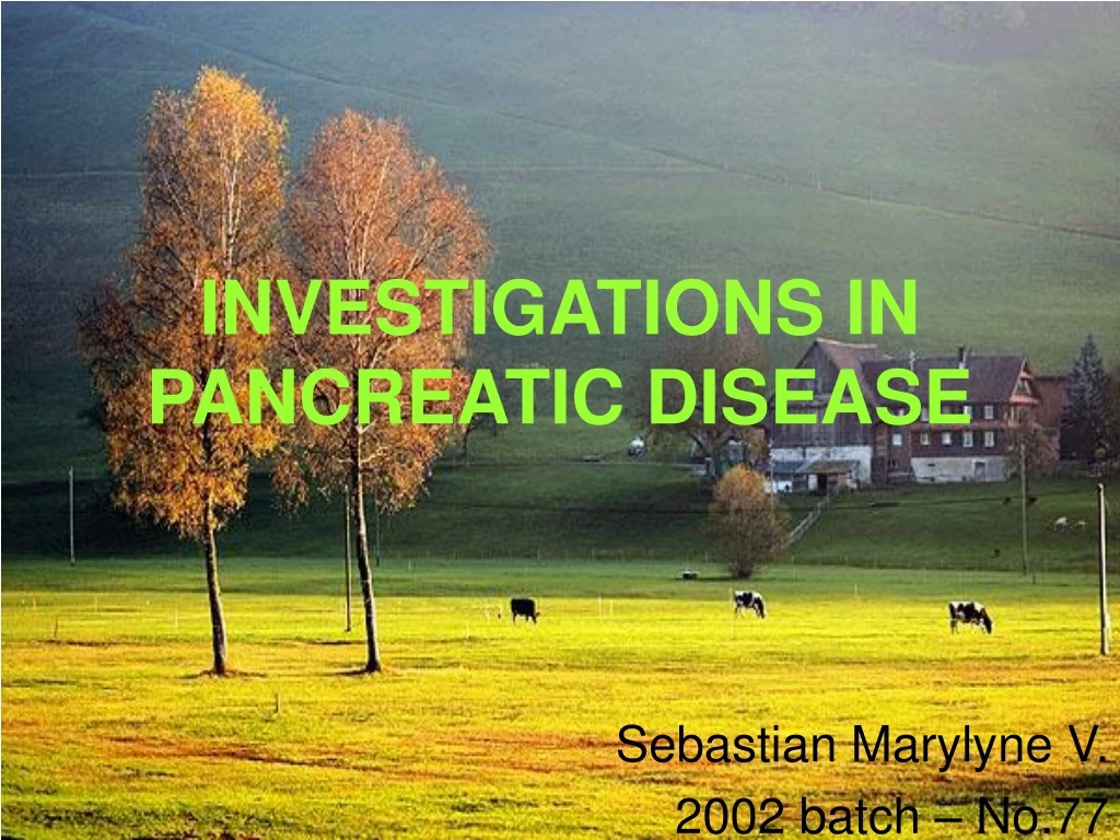
INVESTIGATIONS IN PANCREATIC DISEASE. Sebastian Marylyne V. 2002 batch – No.77. LABORATORY INVESTIGATIONS SPECIFIC INVESTIGATIONS. LABORATORY INVESTIGATIONS. BLOOD TESTS : Acute pancreatitis Haematocrit BUN Serum Creatinine Haemoglobin Serum electrolytes – usually normal

E N D
INVESTIGATIONS IN PANCREATIC DISEASE Sebastian Marylyne V. 2002 batch – No.77
LABORATORY INVESTIGATIONS • SPECIFIC INVESTIGATIONS
LABORATORY INVESTIGATIONS • BLOOD TESTS: Acute pancreatitis Haematocrit BUN Serum Creatinine Haemoglobin Serum electrolytes – usually normal WBC Count - ,shift to left
Blood glucose – elevated in cases of associated Diabetes Mellitus, also because of increased release of glucagon and catecholamines and decreased insulin release. • Hypertriglyceridemia – alcohol-induced • Hypocalcemia – saponification - unresponsive to PTH
Hyperbilirubinemia - inflamed pancreas - biliary tract stones - nonobstructive cholestasis • Thrombocytopenia • Fibrin Degradation Products • Fibrinogen – decreased • Prothrombin Time – prolonged • APTT - prolonged
Serum Amylase Serum Lipase Serum Trypsin and Elastase Activation Peptides Met albumin Inflammatory mediators and acute phase reactants
Carcinoma Pancreas • Elevated Bilirubin and ALP – obstructive jaundice • Serum Markers – CEA and CA 19-9 elevated in advanced disease, very high values indicate unresectable and metastatic disease
Other Pancreatic Neoplasms • Serum Somatostatin • Serum Glucagon • Serum VIP >500pg/ml in the presence of secretory diarrhea - Plasma pancreatic polypeptide levels may also be elevated • Serum Gastrin Fasting hypergastrinemia >200pg/ml Gastric acid studies Secretin provocative test
Insulinoma – Whipples’ triad ** Diagnostic test of choice Serum glucose < 45mg/dl Insulin >5 micro units/ml Insulin: Glucose > 0.3 Proinsulin: Insulin >25% Elevated C-Peptide
SPECIFIC INVESTIGATIONS • ESTIMATION OF PANCREATIC ENZYMES IN BODY FLUIDS • TESTS OF EXOCRINE PANCREATIC FUNCTION • IMAGING INVESTIGATIONS
Serum amylase Acute pancreatitis • Values >3 times normal clinch the diagnosis • Usually elevated within 24 hours, returns to normal within 3 to 5 days unless there is extensive necrosis, incomplete ductal obstruction or pseudo cyst formation
Normal values may be obtained if; • There is a delay ( 2-5 days) before blood samples are obtained • The underlying disorder is chronic rather than acute pancreatitis • Hypertriglyceridemia is present • Overwhelming necrosis • Two types of amylase - P-type - S-type
P-type elevated in -Duodenal ulcer perforation -Gangrenous cholecystitis -Small bowel obstruction -Acute intra-abdominal inflammatory conditions S-type elevated in; -Salivary tumours -Liver disease -Chronic sialadenitis -Ovarian tumours
Isoamylase: -represents about 35 to 45% of the total serum amylase -does not return as rapidly to normal as serum amylase in acute pancreatitis. -Its appearance in blood is also taken as a sign of pseudo pancreatic cyst wall maturity • Urinary amylase remains elevated longer, clearance of >5000 IU/24 hrs is abnormal
Serum Lipase – turbidometric assay - Now thought to be the single best enzyme to measure for the diagnosis of acute pancreatitis • Serum Trypsinogen : radioimmunoassay Acute pancreatitis Chronic pancreatitis with steatorrhea Normal values 28 to 58 ng / ml Normal levels seen in Chronic pancreatitis without steatorrhea and in steatorrhea with normal pancreatic function Elevated in renal failure
Pancreatic Polypeptide Confined to pancreas Release stimulated by nutrients and hormones, in parallel to enzyme secretion Decreased levels seen in chronic pancreatitis Fasting levels >125pg/ml argues against chronic pancreatitis and pancreatic cancer Increased levels seen in pancreatic endocrine tumours
DIRECT TESTS • Direct stimulation of pancreas done and duodenal contents aspirated and analysed • Secretin test – based on the principle that the pancreatic secretory response is directly related to the functional mass of pancreatic tissue Secretin i.v. dose 1 Clinical Unit / Kilogram Normal values; 1). Volume output >2.0 ml/kg/hour 2). Bicarbonate conc. > 80 meq/L 3). Bicarbonate output >10 meq/L in one hour
Secretin test measures the secretory capacity of ductular epithelium Abnormal test only means chronic damage is present, does not differentiate between chronic pancreatitis and carcinoma
Combined Secretin-CCK test permits measurement of pancreatic amylase, lipase, trypsin and chymotrypsin. Frank exocrine pancreatic insufficiency-overall reduction Lesser degrees- dissociation between secretin test results and fecal fat excretion.Only small amounts of enzymes necessaryfor digestion.
INDIRECT TESTS • Lundh test meal Test meal Increased release of CCK Increased enzyme output Trypsin concentration measured False negatives - delayed gastric emptying False positives - choledocholithiasis -primary mucosal disease of the gut Disadvantage – it does not measure secretory capacity
Pancreolauryl test Substrate used is Fluorescein Dilaurate which is hydrolysed by a specific pancreatic aryl-esterase after oral ingestion and fluorescein liberated into the intestinal lumen. It is absorbed by the intestinal mucosa and then excreted by the kidneys. Excretion in urine detected by spectrophotometry
Careful interpretation required in; • post-gastrectomy patients • extensive inflammatory disease • gastrointestinal or hepatobiliary disease that markedly alters g.i. transit and absorptive capacity
MEASUREMENT OF INTRALUMINAL DIGESTION PRODUCTS a). Microscopic examination of stool for undigested meat fibres and fat b). Quantitative stool fat determination c). Faecal Nitrogen
MEASUREMENT OF PANCREATIC ENZYMES IN FAECES • Faecal Chymotrypsin and Elastase Decreased elastase seen in chronic pancreatitis and cystic fibrosis
DUAL-LABELLED SCHILLING TEST • Intrinsic Factor (57Co) and Hog R protein (58Co) given together. Protease needed to cleave R protein. Ratio of labelled Cobalamin excreted in urine is an index of exocrine dysfunction
BREATH TESTS • Measure 14CO2 or13CO2 in breath after the ingestion of a labelled substance - Triolein - Cholesteryl octanoate - Mixed triglyceride ( Distearyl, octanoyl glycerol ) - Starch
PLAIN RADIOGRAPHY • Acute pancreatitis “Sentinel-loop” sign Dilated isolated loop adjacent to the pancreas, Usually jejunum
“Colon Cut-Off” sign A gas-filled descending colon that stops abruptly in the middle or left transverse colon
Generalized ileus with air-fluid levels • Retroperitoneal gas-bubbles – gas-forming organisms • Pancreatic calcification- chronic pancreatitis • Renal “halo” sign • Radio-opaque gall-stones – biliary pancreatitis • Basal atelectasis • Splinting of diaphragm
Pleural effusions Most commonly on the left side
Chronic Pancreatitis Extensive pancreatic calcification
GASTROINTESTINAL BARIUM SERIES • Widening of the duodenal C-loop • Antral Pad sign • Rose- thorning of duodenum • Frostberg’s Inverted-3 sign Inverted-3 sign
ULTRASONOGRAPHY • Often the initial investigation for most patients with suspected pancreatic disease • Normal – comma-shaped, in front of aorta and its branches, IVC. • Acute pancreatitis - pseudocysts - edematous, swollen pancreas - gall stones / dilated bile ducts - peripancreatic fluid collections - calcification • Intraoperative ultrasonography
Chronic Pancreatitis - Calcification
Pancreatic carcinoma - distortion of local landmarks - mass lesions of >3 cm are detected as localized, echo- free lesions
CT SCAN • The best imaging study for evaluation of a suspected chronic pancreatic disorder and for the complications of acute and chronic pancreatitis • Useful for guiding percutaneous pancreatic biopsy and cyst aspiration or drainage • Normal - irregular, lobulated, soft tissue density organ lying in the retroperitoneum
Lesions are characterised by ; - enlargement of the pancreatic outline - distortion of pancreatic contour - fluid filling that has a different attenuation co-efficient compared to the normal pancreas • It may be difficult to distinguish between neoplastic and inflammatory lesions
Acute pancreatitis • normal pancreas • pancreatic edema • abscess • phlegmon • pseudocysts
Oral water-soluble contrast agents may be used-enable more precise delineation • Dynamic CT- rapid i.v. administration of contrast. Useful in estimating the degree of pancreatic necrosis • Spiral (helical CT) provides clear images more rapidly and negates artefact caused by movement