Download
1 / 9
90 likes | 144 Views
3D Bioprinting For Cartilage. Lillian Margolis Biomedical Engineering October 21, 2015. Introduction. Tissue Engineering Study of growth of connective tissue Repair or replace tissue Cartilage Connective tissue Avascular Three Types: Hyaline, Elastic, and Fibrous

E N D
3D Bioprinting For Cartilage Lillian Margolis Biomedical Engineering October 21, 2015
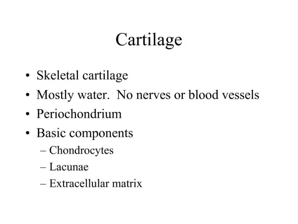
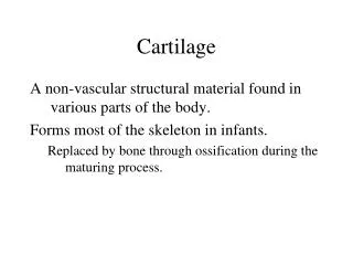
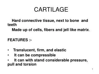
Introduction • Tissue Engineering • Study of growth of connective tissue • Repair or replace tissue • Cartilage • Connective tissue • Avascular • Three Types: Hyaline, Elastic, and Fibrous • 3D Bioprinting: scaffolds and bio-ink [1] [2]
Methods • Scaffolds: three dimensional polymer mold that guides the tissue as it cultures and grows • Mesenchymal stem cells • Chemical cues to mimic the original tissue [3] [3]
Methods • 3D Bioprinting: directly repair or recreate cartilage and integrates with original cartilage • Sizes of tissues printed: under 400 micrometers • 5 options • Extrusion • Laser • Inkjet • Thermal Inkjet • Piezoelectric Inkjet [4] [5]
Studies • Thermal Inkjet Study (Human) • Layer by layer, articular cartilage, and polyethylene glycol dimethacrylate • 4mm diameter, thickness of 2mm, nominal 0.23 microliters bioink • ~1140 chondrocytes • Each layer printed and photopolymerized, 18micrometers thick • 2 mins total printing time • Printed cartilage with 3d biopaper had higher levels of glycosaminoglycan (GAG) content than cartilage printed without • Result: Importance of direct cartilage repair; success in placement of individual cells, preserving cell viability, maintaining chondrogenic phenotype, and integrating with original tissue tissue [5]
Studies • 4 Bioinks: Ink9010, Ink8020, Ink7030, and Ink6040 • Printed small grids with the four different bioinks and crossed-linked them with CaCl2 for 10 mins • Compression testing and shape fidelity testing • The different ink compositions can be used for different printing depending on mechanical properties [6] [6] [6]
Conclusion • Thermal Inkjet Bioprinting • Print both soft and hard tissue • Best option for repairing cartilage • Ink8020 is most suitable bioink for printing • Future • Optimizing scaffolds • Targeted Drug Therapy • Gene Transfection [YW]
Questions? [1]
Resources Besides the Ones in the Abstract • (n.d.). Retrieved October 17, 2015, from http://www.millerplace.k12.ny.us/webpages/lmiller/photos/636532/large23_Cartilage Types.jpg • What is Tissue Engineering. (n.d.). Retrieved October 17, 2015, from http://www.rpi.edu/dept/chem-eng/Biotech-Environ/Projects00/tissue/What is Tissue Engineering.htm • Camarero-Espinosa, S., Rothen-Rutishauser, B., Weder, C., & Foster, E. (2015). Directed cell growth in multi-zonal scaffolds for cartilage tissue engineering. Biomaterials, 42-52. doi:10.1016/j.biomaterials.2015.09.033 • Murphy, S., & Atala, A. (2014). 3D bioprinting of tissues and organs. Nat Biotechnol Nature Biotechnology, 773-785. doi:10.1038/nbt.2958 • Gao, G., & Cui, X. (2015). Three-dimensional bioprinting in tissue engineering and regenerative medicine. Biotechnology Letters, 1-9. doi:10.1007/s10529-015-1975-1 • Markstedt, K., Mantas, A., Tournier, I., Ávila, H., Hägg, D., & Gatenholm, P. (2015). 3D Bioprinting Human Chondrocytes with Nanocellulose–Alginate Bioink for Cartilage Tissue Engineering Applications. BioMacroMolecules, 1489-1496. doi:10.1021/acs.biomac.5b00188 • Digital image. University of Rhode Island. N.p., n.d. Web. <http://www.uri.edu/news/releases/html/images/rhody.jpg>.