Download
1 / 9
90 likes | 220 Views
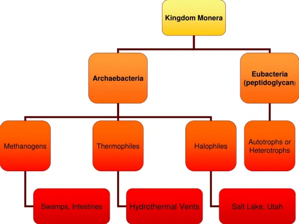
Group 3a Alessandro Borgia Juliane Winkler. FRAP Measure the intracellular (HeLa) diffusion of: EGF (membrane bound) GFP in cytosol. FRAP: membrane bound EGF-receptor. TIME (AFTER BLEACHING). t=100 ms. t=1000 ms. t=1500 ms. t=3000 ms. t=0. FRAP: membrane bound EGF receptor. ROI.

E N D
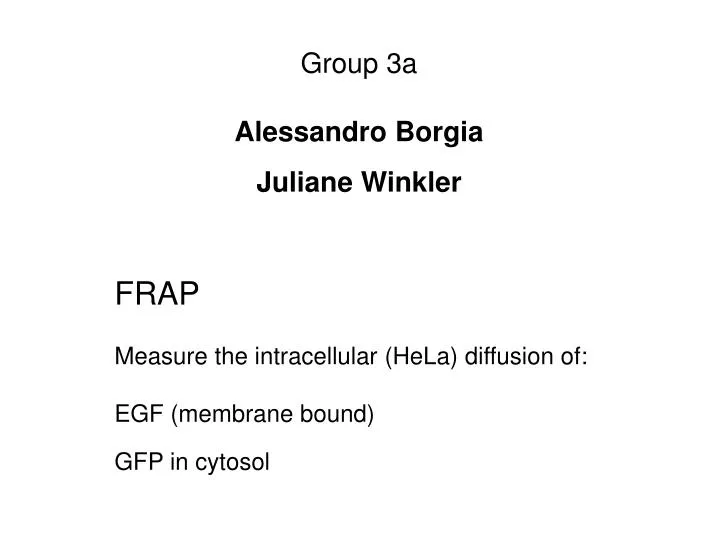
Group 3a Alessandro Borgia Juliane Winkler FRAP Measure the intracellular (HeLa) diffusion of: EGF (membrane bound) GFP in cytosol
FRAP: membrane bound EGF-receptor TIME (AFTER BLEACHING) t=100 ms t=1000 ms t=1500 ms t=3000 ms t=0
FRAP: membrane bound EGF receptor ROI whole cell intensity (cell) mean fluorescence intensity Background (BG) Time (sec)
cell ROI background mean fluorescence intensity Time (sec) EGF-receptor the average intensity of the prebleach image is set to 1 FLUORESCENCE RECOVERY CURVE
Cytosolic GFP PREBLEACH TIME (AFTER BLEACHING) = 130 ms TIME (AB) = 195 ms TIME (AB) = 455 ms
TROUBLESHOOTING (or troubles shooting at us, you decide...) 1ST BLEACHING - SIGNAL POOR ENOUGH LASER POWER ADJUSTED TO MAX DATA STILL VERY NOISY - HOW TO IMPROVE IT? HIGHTEN PHOTOMOLTIPLICATION OR GAIN INCREASE SPOT AREA INCREASES OVERALL SIGNAL INTENSITY • BUT IT TAKES LONGER TO FULLY RECOVER THE FLUO LONGER EXPOSURE TO IMAGING LASER • HIGHER PHOTOBLEACHING OF THE WHOLE CELL FROM IMAGING LASER DID NOT WORK OPTIMIZATION OF THE ACQUISITION ALTOGETHER: REDUCE SCANNING AREA („CLIPPING“) ADJUST BLEACHING TIME TO TRIGGER THE BLEACHING WHEN THE SCANNER IS VERY CLOSE TO THE BLEACHED SPOT INCREASE PIXEL SIZE AND TRIED TO SCAN BIDIRECTIONALLY - LOSE RESOLUTION! FOR CYTOSOL ONLY OPEN THE PINHOLE FULLY - INCREASES SIGNAL AND IS LESS SENSITIVE TO MOTION ALONG Z-AXIS (NO DEFOCUSING PROBLEMS).
THANKS TO: Stefan Terjung Christian Tischer YOU ALL