Download
1 / 56
570 likes | 804 Views
LESSON 8. CARDIAC EMERGENCIES AND CPR. Introduction. Basic cardiac life support needed for patient whose breathing or heart has stopped Ventilations are given to oxygenate blood when breathing is inadequate or has stopped

E N D
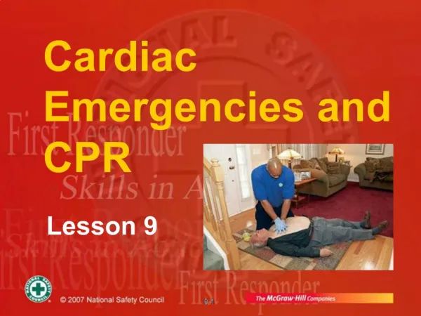
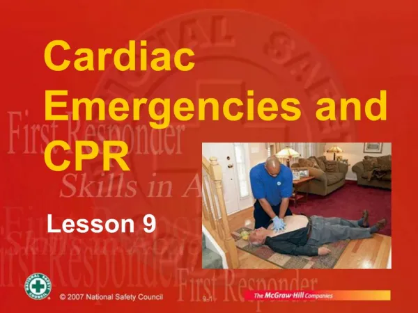
LESSON 8 CARDIAC EMERGENCIES AND CPR
Introduction • Basic cardiac life support needed for patient whose breathing or heart has stopped • Ventilations are given to oxygenate blood when breathing is inadequate or has stopped • If heart has stopped, chest compressions are given to circulate blood to vital organs • Ventilation combined with chest compressions is called CPR • CPR is commonly given to patients in cardiac arrest as a result of heart attack
Review of Circulatory System Circulatory system consists of: • Heart • Blood • Blood vessels
Cardiovascular System: Primary Functions • Transports blood to lungs • Delivers carbon dioxide and picks up oxygen • Transports oxygen and nutrients to all parts of body • Helps regulate body temperature • Helps maintain body’s fluid balance
Anatomy and Physiology of the Heart • Ventricles pump blood through two loops or cycles in body • Right ventricle pumps blood to lungs to pick up oxygen and release carbon dioxide • Blood returns to left atrium and then flows into left ventricle • Left ventricle pumps oxygenated blood through arteries to all areas of body, and to pick up carbon dioxide • Blood returns through veins to right atrium, to be pumped again to lungs • Within heart, valves prevent back flow of blood so that it moves only in one direction through these cycles
Heart Muscle • Heart is composed of a unique type of muscle (myocardium) that contracts to make pumping action
Heart Muscle (continued) • Contractions are controlled by electrical signals under nervous system control
Arteries • Carotid arteries: major arteries passing through neck to head • Femoral arteries:major arteries to legs passing through thigh • Brachial arteries:in upper arm • Radial arteries:major arteries of lower arm • Arteries are generally deeper in body than veins and more protected • Arterial blood is oxygenated, bright red and under pressure
Pulse • When left ventricle contracts, wave of blood is sent through arteries causing pulsing blood pressure changes in arteries that can be palpated in certain body locations • A pulse can be felt anywhere an artery passes near skin surface and over a bone • Palpate carotid pulse on either side of neck
Pulse(continued) • Palpate femoral pulse in crease between abdomen and thigh • Palpate radial pulse on the palm side of wrist proximal to base of thumb • Palpate brachial pulse on the inside of arm between elbow and shoulder
Capillaries • Arteries progressively branch into smaller vessels that eventually reach capillaries • Capillaries are very small blood vessels connecting arteries with veins throughout body • Capillaries have thin walls through which oxygen and carbon dioxide are exchanged with body cells
Veins • From capillaries, blood drains back to heart through extensive system of veins • Venous blood is dark red, deoxygenated and under less pressure than arterial blood • Blood flows more evenly through veins, which don’t have a pulse • Veins have valves that prevent blood backflow
Heart Rate • Heart rate, measured as pulse, is affected by many factors • With exercise, fever or emotional excitement, heart rate increases to meet body’s greater need for oxygen • Various injuries and illnesses may either increase or decrease heart rate
Circulatory System:Emergencies • Any condition that affects respiration – reduces ability to deliver oxygen • Severe bleeding – shock • Stroke – reduces blood flow to brain • Heart conditions – reduce tissue oxygenation
Circulatory System: Emergencies (continued) • Heart attack – can lead to cardiac arrest • Ventricular fibrillation – heart muscle flutters rather than pumping blood
Cardiac Arrest • Heart may stop (cardiac arrest) as a result of heart attack • Brain damage begins 4-6 minutes after cardiac arrest • Brain damage becomes irreversible in 8-10 minutes • Dysrhythmia, an abnormal heartbeat, may also reduce heart’s pumping effectiveness
Causes of Cardiac Arrest • Heart attack • Drowning • Suffocation • Stroke • Allergic reaction • Any condition causing respiratory arrest • Diabetic emergency • Prolonged seizures • Drug overdose • Electric shock • Certain injuries
Call First vs. Call Fast • Call First (to get AED on the way) • If alone with adult found unresponsive and not breathing normally • If alone with patient of any age seen to collapse suddenly • Call Fast • If alone with child found unresponsive and not breathing normally • If alone with patient in cardiac arrest because of asphyxial arrest
CPR • CPR helps keep patient alive by circulating some oxygenated blood to vital organs • Artificial ventilation moves oxygen into lungs where it is picked up by blood • Externalcompressions on sternum increase pressure inside chest, moving some blood to brain/other tissues
CPR (continued) • Blood circulation resulting from chest compressions not as strong as circulation from heartbeat • Can help keep brain and other tissues alive until normal heart rhythm restored
CPR (continued) • Often electric shock from AED is needed to restore a heartbeat and CPR can keep patient viable until then • CPR effective only for a short time • CPR should be started as soon as possible • In some instances, the heart may start again spontaneously with CPR
CPR Saves Lives • CPR and defibrillation within 3-5 minutes can save over 50% of cardiac arrest patients • CPR followed by AED saves thousands of lives each year • In most cases CPR helps keep patient alive until EMS or AED arrives
General Technique of CPR • If unresponsive, not breathing normally and no pulse, start chest compressions • Find the correct hand position • 2 hands for adults • 1 or 2 hands for child • 2 fingers for infant
General Technique of CPR (continued) • Compress chest hard and fast at a rate of at least 100 compressions/minute • Adult = at least two inches deep • Infant/child = at least one-third chest depth • Let chest return to normal height between compressions
General Technique of CPR (continued) • If alone, alternate 30 chest compressions and two ventilations for any age patient • In two-rescuer CPR for infant or child, alternate 15 compressions and two ventilations • Use chest-encircling method in infant • Give each ventilation over one second • Follow local protocol regarding oxygen
Factors That Decrease Effectiveness of Chest Compressions • Compressions that are too shallow • A compression rate that is too slow • Not allowing the chest to recoil fully • Frequent interruptions of compressions
Skill: 1-Rescuer CPR • Determine that the patient is not breathing normally and has no pulse
Give 30 chest compressions at rate of at least 100 per minute Then give two ventilations Continue cycles of compressions and ventilations
Continue CPR until: • Patient is breathing normally and has a pulse • AED brought to scene and ready to use • Personnel with more training arrive and take over
If patient starts breathing normally and has a pulse but is unresponsive, put patient in recovery position and monitor breathing and pulse • When an AED arrives, start AED sequence for patient who is not breathing normally and has no pulse
Chest Compressions Alert • Be careful with your hand position • For adults and children, keep your fingers off patient’s chest • Do not give compressions over bottom tip of breastbone
Chest Compressions Alert (continued) • When compressing, keep elbows straight and hands in contact with patient’s chest at all times
Chest Compressions Alert (continued) • Compress chest hard and fast, but let chest recoil completely between compressions • Minimize amount of time used giving ventilations between sets of compressions
Problems with CPR Technique • CPR often ineffective because of poor technique • Compressions not delivered steadily and constantly during resuscitation efforts • Often compressions are too shallow, resulting in ineffective blood flow • Only good-quality CPR improves chances of survival
Chest Compressions for Bradycardia in Child • Infant or child being given rescue breaths or oxygen may have a pulse but still inadequate perfusion • If pulse <60 beats/minute and infant or child has signs of poor perfusion, provide CPR
2-Rescuer CPR for Adults and Children • Minimizes time between rescue breaths and compressions • CPR becomes more effective • Can more quickly set up AED • Reduces rescuer fatigue
2-Rescuer CPR • Performed in cycles of 30:2 for adult (15:2 for infant or child) • One rescuer provides breaths, second rescuer gives chest compressions • Rescuers switch positions every two minutes • Change done after full CPR cycle • Accomplish change in <5 seconds
2-Rescuer CPR (continued) • If AED present, one rescuer gives CPR while the other sets up unit • If unit advises CPR, rescuers resumeCPR together
2-Rescuer CPR (continued) • If you are assisting another trained rescuer who places an advanced airway: • Chest compressions given continually at rate of at least 100 per minute • No pauses for ventilations • Give ventilations with bag mask at rate of 8-10 breaths/minute
Transitioning from 1-Rescuer CPR to 2-Rescuer CPR • Second rescuer moves into position on other side to prepare to take over chest compressions • First rescuer completes a cycle of compressions and ventilations • While first rescuer gives ventilations, second rescuer finds correct hand position and begins compressions after second ventilation
Differences in 2-Rescuer Training • If EMRstarted CPR, arriving second rescuer may have a higher level of training • Rescuer with greater training determines how CPR should best be continued
Rescuer 1 checks for unresponsiveness, normal breathing, and pulse • Rescuer 2 locates site for chest compressions
If no pulse, rescuer 2 gives 30 compressions for adult (15 for child) at rate of at least100/minute
Continue cycles of 30:2 for adults (15:2 for child) • After 5 cycles (~2 minutes), quickly switch positions
Continue CPR until: • Patient is breathing normally and has a pulse • AED brought to scene and readyto use • Help arrives and takes over • If patient starts breathing normally and has pulse but is unresponsive, put in HAINES recovery position and monitor breathing and pulse • If AED brought to scene, start AED sequence
Infant Two-Rescuer CPR • Uses different hand position • Place thumbs of both hands on sternum just below the nipple line while fingers encircle chest • Compress breastbone with both thumbs • Same rate and depth as usual