Download
1 / 28
380 likes | 989 Views
Gram-Negative Rods Related to Respiratory Tract. By: Lecturer Dr. Thanaa Rasheed. Outline. Haemophilus influenzae , Bordetella pertussis , Legionella pneumophila . Important Properties Diseases and Pathogenesis, Laboratory Diagnosis Treatment and Prevention.

E N D
Gram-Negative Rods Related to Respiratory Tract By: Lecturer Dr. ThanaaRasheed
Outline • Haemophilusinfluenzae, • Bordetellapertussis, • Legionellapneumophila . Important Properties Diseases and Pathogenesis, Laboratory Diagnosis Treatment and Prevention
There are three medically important gram-negative rods : • Haemophilusinfluenzae, • Bordetellapertussis, • Legionellapneumophila . • H. influenzaeand B. pertussisare found only in humans, whereas L. pneumophilais found primarily in environmental water sources.
Haemophilusinfluenzae • Diseases • H. influenzae: Meningitis in young children. • Otitismedia, sinusitis, conjunctivitis, epiglottitis and sepsis in children. • Pneumonia in adults, particularly in those with chronic obstructive lung disease. Haemophilusducreyi, the agent of chancroid,
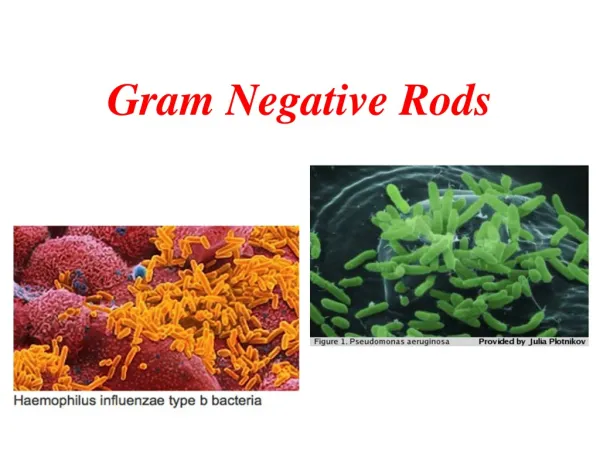
Important Properties • H. influenzae is a small gram-negative rod (coccobacillus) with a polysaccharide capsule. • Serologic typing is based on the antigenicity of the capsular polysaccharide. • Six serotypes, type b causes most of the severe, invasive diseases, such as meningitis and sepsis. • Unencapsulated and therefore untypeable strains can also cause disease, especially diseases of the upper respiratory tract such as sinusitis and otitis media, but are usually noninvasive. • H. influenzae infects only humans; there is no animal reservoir.
Pathogenesis • It enters the body through the upper respiratory tract, resulting in either asymptomatic colonization or infections. • The organism produces an IgA protease that degrades secretoryIgA, thus facilitating attachment to the respiratory mucosa. • The organism can enter the bloodstream (bacteremia) and spread to the meninges. • Meningitis is caused primarily 95% by of which possess the type b capsule, Hib. • The nonencapsulated strains are frequently involved in otitis media, sinusitis, and pneumonia. • Virulence factor involves the antiphagocytic capsule and endotoxin • Noexotoxin is produced.
Clinical Findings • Most infections occur in children between the ages of 6 months and 6 years, with a peak in the age group from 6 months to 1 year. • The rapid onset of fever, headache, and stiff neck along with drowsiness. • Sinusitis and otitis media cause pain in the affected area, opacification of the infected sinus, and redness with bulging of the tympanic membrane. • Septic arthritis, facial cellulitis, and sepsis in splenectomized patients. • Rarely, epiglottitis, which can obstruct the airway. A swollen "cherry-red" epiglottis is seen. • Pneumonia in elderly adults, especially those with chronic respiratory disease, can be caused by untypeable strains of H. influenzae.
Acutely inflamed epiglottis associated with Haemophilus influenza type B Facial cellulitis
Laboratory Diagnosis • Isolation of H. influenzaeon chocolate agar enriched with two growth factors required for bacterial respiration, for adequate energy production namely, factor X (a heme compound) and factor V (NAD). • Other species such as Haemophilusparainfluenzae, do not require both factors. • Biochemical tests • The capsular swelling (quellung) reaction. • Fluorescent-antibody staining (encapsulated) and counterimmunoelectrophoresis or latex agglutination tests, which detect the capsular polysaccharide.
Treatment • The treatment of choice for meningitis or other serious systemic infections caused by H. influenzae is ceftriaxone. • From 20% to 30% of H. influenzae type b isolates produce a β-lactamase that degrades ampicillin but not ceftriaxone. • It is important to institute antibiotic treatment promptly, because the incidence of neurologic sequelae, e.g., subdural empyema, is high. • Untreated H. influenzae meningitis has a fatality rate of approximately 90%. • H. influenzae upper respiratory tract infections are treated with either amoxicillin-clavulanate or trimethoprim-sulfamethoxazole.
Prevention • Vaccine contains the capsular polysaccharide of H. influenzae type b conjugated to diphtheria toxoid or other carrier protein effective in young children. • It is given between the ages of 2 and 15 months. This vaccine is reduced the incidence of meningitis by approximately 90% in immunized children. • Meningitis in close contacts of the patient can be prevented by rifampin. • Rifampin is used because it is secreted in the saliva to a greater extent than ampicillin. • Rifampin decreases respiratory carriage of the organism, thereby reducing transmission.
Bordetella • B. pertussis causes whooping cough (pertussis). • Important Properties • B. pertussis is a small, coccobacillary, encapsulated gram-negative rod.
Pathogenesis & Epidemiology • B. pertussis, a pathogen only for humans • It is transmitted by airborne droplets produced during the severe coughing episodes. • The organisms attach to the ciliated epithelium of the upper respiratory tract but do not invade the underlying tissue. • Decreased cilia activity followed by death of the ciliated epithelial cells. • Pertussis is a highly contagious disease that occurs primarily in infants and young children and has a worldwide distribution.
Virulence factors play a role in the pathogenesis: • Attachment of the organism to the cilia of the epithelial cells is mediated by a protein on the pili called filamentous hemagglutinin • Pertussis toxin • It is an A-B subunit toxin, B mediates its binding to receptors on the surface of respiratory tract epithelial cells.. • A subunit stimulates adenylatecyclase by catalyzing the addition of adenosine diphosphate ribose—a process called ADP-ribosylation—to the inhibitory subunit of the G protein complex (Gi protein). • This results in prolonged stimulation of adenylatecyclase and a consequent rise in cyclic adenosine monophosphate (AMP) and in cyclic AMP-dependent protein kinaseactivity lead to hypersecretion of mucus.
Pertussis toxin also causes a striking lymphocytosis in the blood of patientsis. 3. The organisms also synthesize adenylatecyclase can inhibit their bactericidal activity of neutrophils. 4. Tracheal cytotoxin is a fragment of the bacterial peptidoglycan that damages ciliated cells of the respiratory tract. It act in concert with endotoxin to induce nitric oxide, which kills the ciliated epithelial cells.
Clinical Progression of Pertussis , Inflammation of respiratory mucosal memb. or death Most infectious, but generally not yet diagnosed
Laboratory Diagnosis • The nasopharyngeal swabs taken during the paroxysmal stage. • Nonmotile • Fastidious and slow-growing • Isolated on modified Bordet-Gengou agar • Requires nicotinamide, charcoal, starch, blood, or albumin to absorb toxic substances • Requires prolonged growth • Identification of the isolated organism by agglutination with specific antiserum or by fluorescent-antibody staining. • Polymerase chain reaction–based tests are highly specific and sensitive . • Serological tests that detect antibody in the patient's serum can be used for diagnosis in those patients.
Treatment • Erythromycinreduces the number of organisms in the throat and decreases the risk of secondary complications but has little influence on the course of the disease because the toxins have already damaged the respiratory mucosa. • Supportive care, e.g., oxygen therapy and suction of mucus, during the paroxysmal stage is important, especially in infants.
Prevention • There are two vaccines: • An acellular vaccine containing purified proteins(genetically inactivated toxoid) from the organism • killed vaccine containing inactivated B. pertussis organisms. The acellular vaccine has fewer side effects than the killed vaccine. • The killed vaccine is no longer used because various side effects, including post vaccine encephalopathy at a rate of about one case per million doses administered. • The pertussis vaccine is usually given combined with diphtheria and tetanus toxoids (DTaP) in three doses • beginning at 2 months of age. 2. A booster at 12 to 15 months of age . • 3. And another at the time of entering school • Booster for those between 10 and 18 years old is recommend.
Legionella • L. pneumophila (and other legionellae) causes pneumonia, both in the community and in hospitalized immunocompromised patients. • Important Properties • Legionellae are gram-negative rods that stain faintly with the standard Gram stain. They do, however, have a gram-negative type of cell wall, and increasing the time of the safranincounterstain enhances visibility. • Legionellae in lung biopsy sections do not stain by the standard hematoxylin-and-eosin (H&E) procedure. • Special methods, such as the Dieterle silver impregnation stain.
4. The organism require a high concentration of iron and cysteine; culture media supplemented with these nutrients will support growth. 5. L. pneumophila causes approximately 90% of pneumonia . There are about 30 other Legionella species that cause pneumonia, but most of the remaining 10% of cases are caused by two species, Legionellamicdadei and Legionellabozemanii.
Pathogenesis & Epidemiology • Legionellae are associated chiefly with environmental water sources such as air conditioners and water-cooling towers. • Outbreaks of pneumonia in hospitals have been attributed to the presence of the organism in water taps, sinks, and showers. • The portal of entry is the respiratory tract, and pathologic changes occur primarily in the lung. • In severe cases, bacteremia occurs accompanied by damage to the vascular endothelium in multiple organs, especially the brain and kidneys. • The major virulence factor of the organism is lipopolysaccharide (endotoxin). • No exotoxins are produced.
6. The typical candidate for legionnaires' disease is an older man who smokes and consumes substantial amounts of alcohol. Patients with AIDS, cancer, or transplants (especially renal transplants) or patients being treated with corticosteroids. 7. Despite airborne transmission of the organism, person-to-person spread does not occur.
Clinical Findings • Vary from a mild influenza like illness to a severe pneumonia accompanied by mental confusion, nonbloody diarrhea, proteinuria, and microscopic hematuria. • Although cough is a prominent symptom, sputum is frequently scanty and nonpurulent. Hyponatremia (serum sodium <130 mEq/L) is an important laboratory finding. • Most cases resolve spontaneously in 7 to 10 days, but in older or immunocompromised patients, the infection can be fatal. • Legionellosis is an atypical pneumonia and must be distinguished from other similar pneumonias such as Mycoplasma pneumonia, viral pneumonia, psittacosis, and Q fever. • Pontiac fever is a mild, flu like form of Legionella infection that does not result in pneumonia.
Laboratory Diagnosis • Sputum Gram stains reveals many neutrophils but no bacteria. • The organism fails to grow on ordinary media in a culture of sputum or blood, but it will grow on charcoal-yeast agar, a special medium supplemented with iron and cysteine. • Diagnosis usually depends on a significant increase in antibody titer in convalescent-phase serum by the indirect immunofluorescence assay. • Detection of L. pneumophila antigens in the urine is a rapid means of making a diagnosis. • If tissue is available, it is possible to demonstrate Legionella antigens in infected lung tissue by using fluorescent-antibody staining. • The cold-agglutinin titer does not rise in Legionella pneumonia, in contrast to pneumonia caused by Mycoplasma.
Treatment • Azithromycin or erythromycin (with or without rifampin). • Certain fluoroquinolones, such as levofloxacin and trovafloxacin, are also drugs of choice. • The organism frequently produces β -lactamase, and so penicillins and cephalosporins are less effective. • Prevention • Prevention involves reducing cigarette and alcohol consumption, • Eliminating aerosols from water sources. • Reducing the incidence of Legionella in hospital water supplies by using high temperatures and hyperchlorination. • There is no vaccine.