Download
1 / 62
620 likes | 1.07k Views
ACUTE CORONARY SYNDROMES: Acute MI and Unstable Angina. Tintinalli Chapter 50 September 20, 2005. Acute Coronary Syndrome (ACS). Ischemic heart disease accounts for 500,000 deaths annually in the U.S. CAD and myocardial ischemia contribute to > 5 million ER visits yearly for chest pain

E N D
ACUTE CORONARY SYNDROMES: Acute MI and Unstable Angina Tintinalli Chapter 50 September 20, 2005
Acute Coronary Syndrome (ACS) • Ischemic heart disease accounts for 500,000 deaths annually in the U.S. • CAD and myocardial ischemia contribute to > 5 million ER visits yearly for chest pain • 15% of pts with chest pain will have acute MI and 25-30% will have unstable angina
ACS • a term used to describe pts with acute CP and other symptoms of myocardial ischemia • During the initial exam, often not possible to determine whether permanent damage to the myocardium has occurred • Only in retrospect after serial ECGs or cardiac markers can the distinction b/w AMI or UA be made
Pathophysiology • ACS is caused by secondary reduction in myocardial blood flow due to • coronary arterial spasm • disruption of atherosclerotic plaques • platelet aggregation or thrombus formation at site of atherosclerotic lesion
Thrombus formation • Atherosclerotic plaque formation occurs through repetitive injury to vessel wall • When plaque ruptures, potent thrombogenic substances are exposed to platelets • These platelets respond by adhesion, activation, and aggregation thus initiating thrombus formation in the coronary vessels
The extent of O2 deprivation and thus clinical presentation of ACS depend on the limitation of O2 delivery by thrombus adhering to fixed, fissured, or eroded plaques
Stable Angina • Ischemia occurs only when activity induces O2 demands beyond the supply restrictions imposed by a partially occluded coronary vessel • occurs at a relatively fixed and predictable point and changes slowly over time • atherosclerotic plaque has not ruptured thus there is little superimposed thrombus
ACS • Atherosclerotic plaque rupture and platelet-rich thrombus develop • Degree and duration of O2 supply-demand mismatch determines whether reversible myocardial ischemia w/o necrosis (unstable angina) or myocardial ischemia w/ necrosis (myocardial infarction)
Clinical Features • Main symptom of ischemic heart disease is chest pain • need to characterize its severity, location, radiation, duration, and quality • ask about associated symptoms: N/V, diaphoresis, dyspnea, lightheadedness, syncope, palpitations
Reproducible chest wall tenderness is not uncommon • Patients with ACS may complain of easy fatigability • Usually an AMI is accompanied by more prolonged and severe chest discomfort and more prominent associated symptoms
Angina Pectoris • Exercise, stress, or cold environment classically precipitates angina • duration of symptoms typically < 10 minutes, occasionally lasting up to 20 minutes • usually improves within 2-5 minutes after rest or nitroglycerin
ACS • Up to 30% of patients with AMI are clinically unrecognized • Some of these patients have had atypical symptoms for which they didn’t pursue medical advice • Worse prognosis for pts who have atypical symptoms at the time of their infarction • women and elderly most likely to have atypical symptoms
Age over 40 male postmenopausal females family history cigarette smoking hypertension High cholesterol truncal obesity sedentary lifestyle diabetes previous cardiac hx Cardiac Risk Factors
Cardiac Risk Factors • Risk factors are modestly predictive of CAD is asymptomatic patients • In the ER, risk factors are poor predictors of cardiac risk for MI or other ACS • In males, only DM and family history are weakly predictive • Cardiac risk factors are not predictive of ACS in female ER chest pain pts
Physical Examination • Not helpful in distinguishing pts with ACS from those with non cardiac etiologies • Pts may appear deceptively will without distress or be uncomfortable, pale, cyanotic, and in respiratory distress.
Vital Signs • Bradycardic rhythms are more common with inferior wall MI • in the setting of anterior wall MI, bradycardia or heart block is very poor prognostic sign • Extremes of blood pressures are associated with worse prognosis
Heart Sounds • S1 and S2 are often diminished due to poor myocardial contractility • S3 is present in 15-20% of pts with AMI • implies a failing myocardium • S4 is common in pts with long standing HTN or myocardial dysfunction • Presence of new systolic murmur is an ominous sign • signifies papillary m. dysfunction, flail leaflet of mitral valve, or VSD
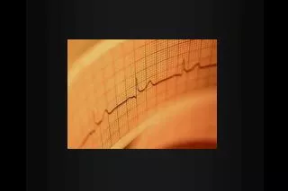
ECG • 12 lead is single best test to identify pts with AMI upon presentation to ER • Current guidelines state that the initial 12 lead ECG must be obtained and interpreted within 10 minutes of patient presentation • Yet ECG has a relatively low sensitivity for detection of AMI
ECG • ST segment is elevated on the initial ECG in approximately 50% of pts with AMI • most other AMI pts will have ST depression and/or T wave inversions • Only 1-5% of pts with AMI have an entirely normal initial ECG
Anteroseptal --> Anterior --> anterolateral --> QS deflections in V1-V3, possibly V4 rS defection in V1, Q waves V2-4 or decr in amplitude of initial R wave in V1-V4 Q waves in V4-6, I, aVL ECG criteria and AMI
Lateral --> inferior --> inferolateral --> true posterior --> right ventricular --> Q waves in I, aVL Q waves II, III, aVF Q waves II, III, aVF, and V5-V6 Initial R waves in V1-V2 >0,04s and R/S ratio > 1 Q waves II, III, aVF & ST elevation rV4 ECG Criteria and AMI
ECG • In distributions previously described: • ST elevation suggests acute transmural injury • ST depression suggests subendocardial ischemia • All inferior wall MI should have right sided ECG • ST elevation in rV4 indicates right ventricular infarction
ECG • Reciprocal ST segment changes predict: • a larger infarct distribution • an increased severity of underlying CAD • more severe pump failure • a higher likelihood of cardiovascular complications • increased mortality
Difficult ECG interpretations • ST elevation in absence of AMI • early repolarization • LVH • pericarditis/myocarditis • Left ventricular aneurysm • Hypertropic cardiomyopathy • hypothermia • ventricular paced rhythms • LBBB
Difficult ECG interpretations • ST depression in absence of ischemia • hypokalemia • digoxin effect • cor pulmonale and right heart strain • early repolarization • LVH • ventricular paced rhythms • LBBB
Difficult ECG interpretations • T wave inversions without ischemia • persistent juvenile pattern • seizures or Stokes Adams syncope • post-tachycardia T wave inversion • post-pacemaker T wave inversion • Intracranial pathology (CNS hemorrhage) • Mitral valve prolapse • pericarditis • primary or secondary myocardial disease
T wave inversion without ischemia • PE or cor pulmonale • spontaneous PTX • myocardial contusion • LVH • ventricular paced rhythms • RBBB • LBBB
AMI and LBBB • In the setting of LBBB, the following are indicative of AMI • 1. ST elevation 1mm or greater and concordant with the QRS complex • 2. ST depression 1mm or more in leads V1, V2, or V3 • 3. ST elevation 5mm or greater and discordant with the QRS complex
Cardiac Enzymes • Serial measurements are more sensitive and accurate than initial single measurement • serum markers have less utility in the diagnosis of UA, only about 50% will have elevated troponins
CK-MB • Most commonly used marker in ACS • a serial rise to above 5 times baseline followed by fall back to baseline is considered diagnostic for AMI • peaks at 12-24 hours, with fall back to baseline in 2-3 days • useful in detecting recurrent infarction after the initial 24-48 hours by noting a repeat elevation in the level
Unstable angina acute coronary ischemia inflammatory heart disease cardiomyopathies circulatory failure & shock DTs Rhabdomyolysis Cardiac surgery skeletal m. trauma dermatomyositis, polymyositis myopathic disorders muscular dystrophy vigorous exercise malignant hyperthermia Ethanol poisoning (chronic) Conditions Associated with Elevated CK-MB
Troponin • Main regulatory protein for the actin-myosin myofibrils • 3 subunits: • inhibitory subunit (Trop I) • tropomyosin binding subunit (Trop T) • calcium binding subunit (Trop C) • Trop I has not been identified in skeletal m. during any stage of develop therefore specific to myocardium
Troponin • Peak level in 12 hours • prolonged elevation for 7 to 10 days before returning to baseline • thus making trop of no use in detecting recurrent infarctions during this time • Rise in serum Trop I or T is considered diagnostic for AMI • Low level elevations in Trop correlate with risk for CV complications in UA, CAD, and renal failure
Myoglobin • Rises within 2-3 hours of symptoms onset • peaks within 4 to 24 hours • more sensitive than CK and CK-MB but not specific for cardiac muscle • there is a high false-positive rate due to its presence in all muscle tissue
Complications of MI • 1. Dysrhythmias and conduction disturbances • 2. Cardiac failure • 3. Mechanical complications • 4. Pericarditis • 5. Right Ventricular Infarction • 6. Other
Dysrhythmias • Occurs in 72-100% of AMI pts treated in coronary care unit • PVCs are common in AMI • occur in >90% of AMI patients • Atrial premature contractions are also common • occur in up to 50% of AMI patients • not associated with increased mortality
Dysrhythmias • Early in AMI, pts often show increased autonomic nervous system activity • sinus brady, AV block, hypotension occur from increased vagal tone • Later, increased sympathetic activity results in incr catecholamine release • thus creates electrical instability: PVCs, Vtach, Vfib, accelerated idioventricular rhythms, AV junctional tachycardia
Dysrhythmias • Hemodynamic consequences of dysrhythmias are dependent on ventricular function • Normal hearts have a loss of 10-20% of left ventricular output when atrial kick is eliminated • Reduced left ventricular compliance can result in 35% reduction in stroke volume when the atrial systole is eliminated
Dysrhythmias • Persistant tachycardia is associated with poor prognosis • due increase myocardial oxygen use • When Vtach occurs late in AMI course, usually associated with transmural infarct and left ventricular dysfunction • induces hemodynamic deterioration • mortality rate approaches 50%
Conduction Disturbances • First degree and Mobitz I (Wenckebach) • more common with inferior AMI • intermittent during the first 72 hrs after infarction • rarely progresses to complete block or pathologic rhythm • Mobitz II • usually associated with anterior AMI • does progress to complete heart block
Conduction Disturbances • Complete Heart Block • occurs in setting of inferior MI • usually progresses from less AV blocks • this form is usually stable & should resolve • Mortality is 15% in absence of RV involvement & increases to 30% when RV is affected • Complete block in setting of anterior MI results in grave prognosis
Conduction Disturbance • New RBBB • occurs in approximately 2% of AMI pts • associated with anteroseptal AMI • associated with increased mortality because often leads to complete AV block
Conduction Disturbance • New LBBB • occurs in 5% of pts with AMI • associated with high mortality • Left posterior hemiblock associated with higher mortality than isolated anterior hemiblock • represents larger area of infarction
Cardiac Failure • 15-20% of AMI pts present in some degree of CHF • More severe the degree of left ventricular dysfunction, the higher the mortality • dependent on the net effect of prior myocardial dysfunction, baseline myocardial hypertrophy, acute myocardial necrosis, & acute reversible dysfunction (“stunned myocardium”)
Cardiac Failure • B-type natriuretic peptide • useful for risk stratification of pts with non ST elevation MI and UA • elevated levels of BNP early in the hospital course predict a worse outcome at 30 days
Mechanical Complicationsof AMI • Sudden decompensation of previously stable AMI pt should raise concern of the “mechanical” complication • Free wall rupture • occurs in 10% of AMI fatalities, usually 1 to 5 days after infarction • rupture of LV free wall usually leads to pericardial tamponade and death (>90% of cases)
Mechanical Complicationsof AMI • NSAIDs, steroids, and late administration of thrombolytics have been linked to an increased likelihood of cardiac rupture • however, studies remain contradictory • LV hypertrophy appears to be protective
Mechanical Complicationsof AMI • Rupture of interventricular septum • is more often detected clinically than ventricular wall rupture • pts have chest pain, dyspnea, sudden appearance of new holosystolic murmur • murmur often associated with palpable thrill and best heard at lower left sternal border • more common in pts with anterior wall MI and pts with extensive (3 vessel) CAD
Mechanical Complicationsof AMI • Papillary Muscle Rupture • occurs in 1% of pts with AMI • more common with inferior wall MI • usually occurs 3 to 5 days after AMI • occurs with a small to modest sized MI • posteromedial m. commonly ruptured • receives blood from only one coronary a. • present with acute dyspnea, increasing CHF, and new holosystolic murmur consistent with mitral regurgitation
Pericarditis • Occurs in 10-20% of post-AMI pts • more common with transmural MI • usually occurs 2-4 days after AMI • Pericardial friction rubs detected more often with inferior wall and right ventricular infarcts • Pericardial effusions may also be present; may take months to resorb