Download
1 / 19
190 likes | 392 Views
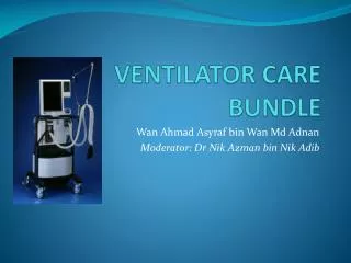
VENTILATOR – ACQUIRED PNEUMONIA By Prof. Adel Salah Professor of Respiratory Medicine Zagazig University . Definition. VAP refers to pneumonia that arises more than 48 hours after intubation of trachea and initiation of M. ventilation .

E N D
VENTILATOR – ACQUIRED PNEUMONIA • By • Prof. Adel Salah • Professor of Respiratory Medicine • Zagazig University
Definition VAP refers to pneumonia that arises more than 48 hours after intubation of trachea and initiation of M. ventilation. • According to the microbial pattern and the clinical outcome, VAP can be divided into: • Early onset VAP. • Late onset VAP.
Determinants of the microbiologic spectrum of VAP: • Prior use of antibiotics. • Duration of time on mechanical ventilation Risk Factors for MDR Pathogens Causing VAP and HAP(Am J Respir Crit Care Med , 2005)
Pathogenesis • Colonization of aero-digestive tract . • Aspiration of infected secretions . • Insertion of an ETT: • comprises the natural barrier. • facilitates pooling and leakage of contaminated secretions. • eliminates the cough reflex. • impair mucociliary clearance. • bacterial biofilm . • injure the epithelial surface . • Other factors: supine position, frequency of ventilator circuit changes, tracheal suctioning, and contaminated respiratory care equipment . • Compromise of the normal host defense mechanisms.
Diagnosis of VAP The clinical pulmonary infection score (CPIS) used for the diagnosis of VAP (Luna et al., 2003) CPIS > 6 is associated with a high likelihood of pneumonia.
Signs and symptoms are very nonspecific and lead to overdiagnosis of pneumonia. So why it is difficult to accurately diagnose VAP? • Fever and leucocytosis. • Increased tracheal secretion. • Oropharyngeal colonization. • Chest radiographic changes. • Blood or pleural fluid culture. How can pneumonia be differentiated from all these other diseases? Confirmation of VAP usually relies on a lower respiratory tract quantitative cultures
What is the rationale for QCs Infection is more likely the higher the density of microorganisms in a particular space. A critical density must be reached in order to cause clinical infection. A certain threshold is therefore used in order to separate true infection from colonization. • B.PSB > 100-1000 CFU/ml • B.BAL > 1000-10000 CFU/ml • Deep endotrahceal aspirates > 105-106 CFU/ml
The most commonly used of QCs techniques • Bronchoscopic: • Bronchoalveolarlavage • Protected specimen brush • Deep tracheal aspirates. • Nonbronchoscopic catheters. • Distal sampling, mini-BAL (e.g., Combicath) • Nonbronchoscopic protected specimen brush Advantages and disadvantages of QCs Is one bronchoscopic technique better than the other?
Role of biomarkers as diagnostic and prognostic markers of VAP • Procalcitonin (PCT): • Low PCT levels (< 0.25 µg/l) in patients with no clinical signs of severe illness suggest safe withdrawal of antibiotics, thereby limiting the use of unnecessary antibiotics. • Alternatively PCT > 0.5 µg/ml strongly recommends antibiotic treatment as it is indicative of active bacterial infection. • PCT was shown to be elevated on an average 2 days prior to the clinical diagnosis of VAP and therefore can be used as an early marker for diagnosis of VAP. The other biomarkers: like natriuretic peptides, copeptin and CRP levels are useful prognostic markers and may be of great help in the risk stratification of patients.
Differential diagnosis • Other diagnoses should be considered in a patients without typical signs and symptoms or those who do not respond to empirical antibiotic therapy. • The differential diagnosis of VAP includes: • congestive heart failure. • Atelectasis. • Acute respiratory distress syndrome (ARDS). • pulmonary embolism with infarction. • chemical pneumonitis from aspiration. • alveolar hemorrhage. • Lung contusion in trauma patient.
Complications • Patients who remain ill after initiation of appropriate antibiotic therapy may have developed complications of pneumonia. • Common complications include : • complicated parapneumonic effusions, frank empyema. • ARDS. • Spreading of infection to other organs. • sepsis, with multiple organ dysfunction, Renal failure hepatic failure, disseminated intravascular coagulation, hemodynamic instability, and coma may all occur in this setting.
Antibiotic treatment of VAP 2 overriding principles • Adequate initial empiric antibiotic therapy. • Avoiding unnecessary antibiotics. Duration of therapy Antibiotic class rotation
Treatment Failure • criteria indicate treatment failure: (usually after 3 days from AB initiation): • failure to improve the PaO2/FiO2 ratio after antibiotic initiation. • persistence of fever or hypothermia plus purulentrespiratory secretions. • worsening of the pulmonary infiltrate by 50%. • development of septic shock or multiple organ failure syndrome. causes • Wrong diagnosis. • Host factors: underlying diseases (e.g., endobronchial malignancy or foreign body), superinfectionswith organisms that are not sensitive to the antibiotics given, or any of a variety of types of immunosuppression. • Bacterial factors : associated with failure of initial therapy include primary or acquired resistance to the initial antibiotic. • Complications of the initial infection: (e.g., empyema, abscess formation).
Prevention strategies • Guard against Bacterial colonization : • Effective hand washing by hospital personnel. • Protective gowns and gloves. • Oral (nonnasal) intubation. • Limiting stress ulcer prophylaxis to high risk critically ill patients such as those who require mechanical ventilation or have a coagulopathy. • Avoiding unnecessary antibiotics. • Adequate initial empiric antibiotic therapy. • Antibiotic class rotation. • Chlorhexidine oral rinse. • Selective digestive decontamination • Probiotics. • Antimicrobial coating of tracheal tubes.
Guard against aspiration of contaminated secretions: • Keeping mechanically ventilated patients semirecumbent. • Reducing excessive use of narcotics. • Continuous subglottic suctioning. • Use of a closed endotracheal suctioning system. • Use of weaning protocols to shorten duration of mechanical ventilation. • Do not routinely change the ventilator circuit. Only change when visibly soiled or malfunctioning. • Consider non invasive ventilation • Drain circuit condensate. • Use only sterile fluid for humidification or nebulization. • Avoiding large gastric volumes. • Avoid patient transport. • Reduce accidental extubations.