Download
1 / 1
10 likes | 103 Views
Combinatorial Selection of Material Specific Peptides Marketa Hnilova 1 , E. Emre Oren 1 , Mustafa Gungormus 1 , Turgay Kacar 1,2 , Candan Tamerler 1,2 and Mehmet Sarikaya 1,2 1 Materials Science and Engineering and GEMSEC, University of Washington, Seattle, WA 98195, USA,

E N D
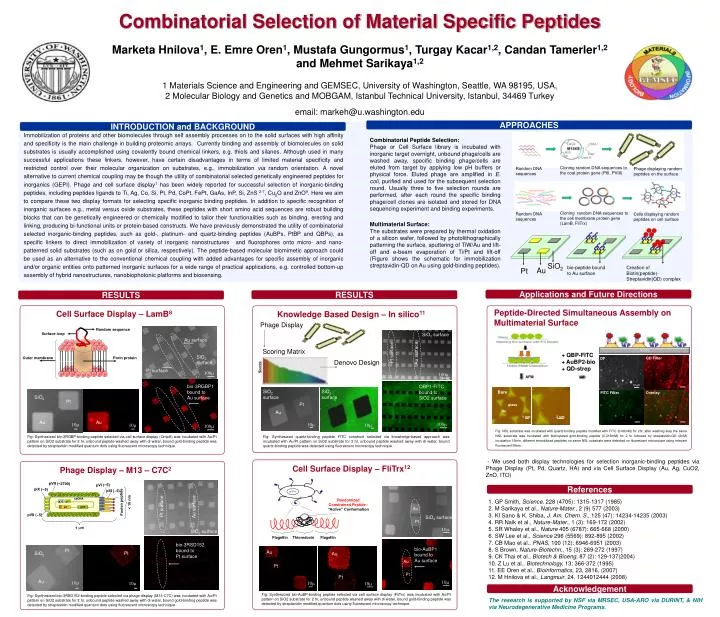
Combinatorial Selection of Material Specific Peptides Marketa Hnilova1, E. Emre Oren1, Mustafa Gungormus1, Turgay Kacar1,2, Candan Tamerler1,2 and Mehmet Sarikaya1,2 1 Materials Science and Engineering and GEMSEC, University of Washington, Seattle, WA 98195, USA, 2 Molecular Biology and Genetics and MOBGAM, Istanbul Technical University, Istanbul, 34469 Turkey email: markeh@u.washington.edu APPROACHES INTRODUCTION and BACKGROUND Immobilization of proteins and other biomolecules through self assembly processes on to the solid surfaces with high affinity and specificity is the main challenge in building proteomic arrays. Currently binding and assembly of biomolecules on solid substrates is usually accomplished using covalently bound chemical linkers, e.g. thiols and silanes. Although used in many successful applications these linkers, however, have certain disadvantages in terms of limited material specificity and restricted control over their molecular organization on substrates, e.g., immobilization via random orientation. A novel alternative to current chemical coupling may be though the utility of combinatorial selected genetically engineered peptides for inorganics (GEPI). Phage and cell surface display1 has been widely reported for successful selection of inorganic-binding peptides, including peptides ligands to Ti, Ag, Co, Si, Pt, Pd, CoPt, FePt, GaAs, InP, Si, ZnS 2-7, Cu2O and ZnO9. Here we aim to compare these two display formats for selecting specific inorganic binding peptides. In addition to specific recognition of inorganic surfaces e.g., metal versus oxide substrates, these peptides with short amino acid sequences are robust building blocks that can be genetically engineered or chemically modified to tailor their functionalities such as binding, erecting and linking, producing bi-functional units or protein-based constructs. We have previously demonstrated the utility of combinatorial selected inorganic-binding peptides, such as gold-, platinum- and quartz-binding peptides (AuBPs, PtBP and QBPs), as specific linkers to direct immobilization of variety of inorganic nanostructures and fluorophores onto micro- and nano-patterned solid substrates (such as on gold or silica, respectively). The peptide-based molecular biomimetic approach could be used as an alternative to the conventional chemical coupling with added advantages for specific assembly of inorganic and/or organic entities onto patterned inorganic surfaces for a wide range of practical applications, e.g. controlled bottom-up assembly of hybrid nanostructures, nanobiophotonic platforms and biosensing. Combinatorial Peptide Selection: Phage or Cell Surface library is incubated with inorganic target overnight, unbound phage/cells are washed away, specific binding phage/cells are eluted from target by applying low pH buffers or physical force. Eluted phage are amplified in E. coli, purified and used for the subsequent selection round. Usually three to five selection rounds are performed, after each round the specific binding phage/cell clones are isolated and stored for DNA sequencing experiment and binding experiments. Cloning random DNA sequences to the coat protein gene (PIII, PVIII) Random DNA sequences Phage displaying random peptides on the surface Cloning random DNA sequences to the cell membrane protein gene (LamB, FliTrx) Random DNA sequences Cells displaying random peptides on cell surface Multimaterial Surface: The substrates were prepared by thermal oxidation of a silicon wafer, followed by photolithographically patterning the surface, sputtering of TiW/Au and lift-off and e-beam evaporation of Ti/Pt and lift-off (Figure shows the schematic for immobilization streptavidin-QD on Au using gold-binding peptides). SiO2 bio-peptide bound to Au surface Creation of Biotin(peptide)-Streptavidin(QD) complex Au Pt Applications and Future Directions RESULTS RESULTS Peptide-Directed Simultaneous Assembly on Multimaterial Surface Cell Surface Display – LamB8 Knowledge Based Design – In silico11 Phage Display Random sequence Surface loop SiO2 surface Au surface Pt surface Au surface Scoring Matrix + QBP-FITC + AuBP2-bio + QD-strep SiO2 surface Outer membrane Porin protein Denovo Design Scores Pt surface QD Filter 100m DF 100m AFM bio-3RGBP1 bound toAu surface QBP1-FITC bound toSiO2 surface SiO2 surface SiO2 surface 10mm Bare SiO2 FITC Filter Overlay Pt Pt glass Au Au 5m 1 um 2 um Au Au 100m 10m 10m 10m 10m 100m Fig: NSL substrate was incubated with quartz-binding peptide modified with FITC (0.06mM) for 2hr, after washing step the same NSL substrate was incubated with biotinylated gold-binding peptide (0.016mM) for 2 hr followed by streptavidin-QD (2nM) incubation 15min, different immobilized peptides on same NSL substrate were detected on fluorescent microscope using relevant fluorescent filters. Fig: Synthesized quartz-binding peptide FITC construct selected via knowledge-based approach was incubated with Au/Pt pattern on SiO2 substrate for 2 hr, unbound peptide washed away with di water, bound quartz-binding peptide was detected using fluorescent microscopy technique. Fig: Synthesized bio-3RGBP-binding peptide selected via cell surface display (OmpA) was incubated with Au/Pt pattern on SiO2 substrate for 2 hr, unbound peptide washed away with di water, bound gold-binding peptide was detected by streptavidin modified quantum dots using fluorescent microscopy technique. - We used both display technologies for selection inorganic-binding peptides via Phage Display (Pt, Pd, Quartz, HA) and via Cell Surface Display (Au, Ag, CuO2, ZnO, ITO) Cell Surface Display – FliTrx12 Phage Display – M13 – C7C2 pVII (~2700) pVI (~5) pIX (~5) pFliTrx pIII (~5) References Fusion peptide pVIII pVIII pVIII pVIII pVIII pVIII pVIII pVIII pVIII pVIII pVIII pVIII pVIII pVIII pVIII Randomized Constrained Peptide - “Active” Conformation ssDNA Au < 10 nm 1. GP Smith, Science, 228 (4705): 1315-1317 (1985) 2. M Sarikaya et al., Nature-Mater., 2 (9) 577 (2003) 3. KI Sano & K. Shiba, J. Am. Chem. S., 125 (47): 14234-14235 (2003) 4. RR Naik et al., Nature-Mater., 1 (3): 169-172 (2002) 5. SR Whaley et al., Nature 405 (6787): 665-668 (2000) 6. SW Lee et al., Science 296 (5569): 892-895 (2002) 7. CB Mao et al., PNAS, 100 (12): 6946-6951 (2003) 8. S Brown, Nature-Biotechn., 15 (3): 269-272 (1997) 9. CK Thai et al., Biotech & Bioeng, 87 (2): 129-137(2004) 10. Z Lu et al., Biotechnology, 13: 366-372 (1995) 11. EE Oren et al., Bioinformatics, 23, 2816, (2007) 12. M Hnilova et al., Langmuir, 24, 1244012444 (2008) gIX gVII Pt surface Au surface gIII gVIII SiO2 surface Pt pVII (~5) pVIII pVIII pVIII pVIII pVIII pVIII pVIII pVIII pVIII pVIII pVIII pVIII pVIII pVIII pVIII 1 m 10m SiO2 surface Flagellin Thioredoxin Flagellin Au bio-3RSD152 bound toPt surface bio-AuBP1 bound toAu surface Pt Au Au Pt SiO2 Au Pt Pt Pt Au 10m 10m 10m 10m 10m Acknowledgement Fig: Synthesized bio-AuBP-binding peptide selected via cell surface display (FliTrx) was incubated with Au/Pt pattern on SiO2 substrate for 2 hr, unbound peptide washed away with di water, bound gold-binding peptide was detected by streptavidin modified quantum dots using fluorescent microscopy technique. Fig: Synthesized bio-3RSD152-binding peptide selected via phage display (M13-C7C) was incubated with Au/Pt pattern on SiO2 substrate for 2 hr, unbound peptide washed away with di water, bound gold-binding peptide was detected by streptavidin modified quantum dots using fluorescent microscopy technique. The research is supported by NSF via MRSEC, USA-ARO via DURINT, & NIH via Neurodegenerative Medicine Programs. GLASS