Download
1 / 30
300 likes | 303 Views
Learn about the importance of proper protein folding and the role of ubiquitination in protein degradation. This lecture covers protein translocation, ER stress, chaperones, and their role in disease.

E N D
Gene regulationLecture No 5: Protein folding and Ubiquitination Dr. Mohamed Kamal Lecturer of Molecular Biology E.mail:mk_saleh1980@yahoo.com
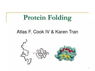
Protein folding Correct protein folding is essential to proper protein function. Protein folding occurs mainly in the ER. The status of protein folding act as a feedback to the protein translation. Signaling pathways emanate from the ER to regulate mRNA translation. These pathways prevent the accumulation of unfolded protein in the ER by decreasing the load, increasing the ER folding capacity, and increasing the degradation of misfolded proteins.
Protein folding *High proportion of proteins are translocated into the ER. *Proteins are translocated into the ER lumen in an unfolded state. *Inhibition of translation initiation serves as an effective means to limit the flow of proteins into the ER .
Protein folding *Translocation of nearly all proteins across the ER membrane occurs co-translationally and is usually directed by an N-terminal signal peptide. *When the signal peptide is formed the signal recognition particle (SRP) binds and stops translational elongation until the ribosome docks at the ER membrane.
Protein folding Upon docking to the ER membrane, the polypeptide is simultaneously synthesized and translocated across the lipid bilayer. This mechanism prevents the polypeptide from improper localization in the cytosol. ER translocon:An aqueous pore formed by the Sec61 complex and the BiP/GRP78 (immunoglobulin-binding protein/glucose-regulated protein).
Protein folding Functions of BiP: 1- It has a peptide-dependent ATPase activity that is used to seal the lumenal side of the aqueous pore to maintain the permeability barrier between the ER and the cytosol when the ribosome is not tightly attached. 2- BiP functions in post-translational translocation to ensure unidirectional translocation of the elongating polypeptide across the ER membrane.
Protein folding ER Capacity: The protein concentration in the ER lumen is 100 mg/ml, it is essential that protein chaperones facilitate protein folding by preventing aggregation of protein folding intermediates and by correcting misfolded proteins. BiP/GRP78 – uses the energy from ATP hydrolysis to facilitate folding by preventing aggregation of proteins within the ER. Quality control: Only those polypeptides that are properly folded and assembled in the ER can transit to the Golgi compartment. Proteins that are misfolded in the ER are retained and eventually translocated back through the ER translocon into the cytosol for degradation by the 26S proteasome.
Protein folding ER Stress: It means overload of proteins in the ER. Activate misfolded proteins unfolded protein response (UPR) i) protein translocation iii) protein folding capacity ii) ERAD
Protein folding UPR is orchestrated by a general attenuation of translation initiation, a selective translation of a small subset of mRNAs encoding adaptive functions, and transcriptional activation of a large set of genes. Cell death (necrosis or apoptosis) Failure of UPR
Protein folding ER sensors: IRE1, PERK and ATF6
Protein folding Combinatorial interactions of ER sensors generate diversity for transcriptional induction of different subsets of UPR-responsive genes. Example: IRE1, PERK and ATF6 Regulate Basic leucine-zipper (bZIP)-containing transcription factors
Protein folding Regulation of UPR: Free BiP BiP Accumulation of unfolded proteins IRE1, PERK or ATF6 Unstressed ER Activate UPR Fig 1 BiP-unfolded proteins binding
Protein Folding and disease Misfolded proteins accumulat in the cell Accumulation of specific proteins cause different diseases. Example:Accumulation of αsynuclein increases lewy bodies in the neurons and causes Parkinson Disease.
Protein Folding and disease Science News Pathway Identified in Human Lymphoma Points Way to New Blood Cancer Treatments ScienceDaily (Nov. 21, 2012) — A pathway called the "Unfolded Protein Response," or UPR, a cell's way of responding to unfolded and misfolded proteins, helps tumor cells escape programmed cell death during the development of lymphoma.
Protein folding Protein folding is a spontinous process. Chaperons: Are a group of proteins playing important role in protein folding. Chaperons safeguard the folding of nascent chains. Most proteins bind chaperons, however it is essential for the correct folding of a limited number of proteins.
Protein folding * Heat shock proteins (Hsp): They are chaperons. * Hsps stabelize the newly formed chains and protect them from their hydrophobic characters. • * Hsp70-bound substrate must be transferred to a chaperonin complex for productive folding.
Protein folding The chaperonins: are large cylindrical protein complexes consisting of two stacked rings of seven to nine subunits each. The chaperonin of the eukaryotic cytosol, termed TRiC or CCT forms a cage-like structure.
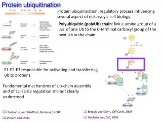
Protein Ubiquitination Protein Degradation Normal protein turnover Elimination of misfolded proteins Protein Degradation Proteasomal by ubiquitination Lysosomal 10–20% of normal protein turnover Aberrations in this pathway provide a variety of pathological phenotypes
Protein Ubiquitination The ubiquitination pathway: The ubiquitin–protein ligase system includes three enzymes, termed E1, E2, and E3, is absolutely necessary, along with adenosine triphosphate (ATP), for the conjugation of ubiquitin to proteins.
Protein Ubiquitination Other functions for ubiquitination - Internalization of proteins (binding to K63) - nuclear export and translocation of proteins into the cytoplasm (MonoUbiquitination)
Protein Ubiquitination The Proteasome: is a protein complex mediates protein degradation and is localised in the nucleus and cytosol
Protein Ubiquitination Proteasome confers trypsin-like activity. Caspase-like effects. It can cleave bonds on the carboxyl side of basic, hydrophobic, or acidic amino acid residues.
Protein Ubiquitination in Cancer * 20S proteasome subunit expression in Cancer * E2 and some E3 ligases high expression in cancer