Download
1 / 75
770 likes | 806 Views
ENTEROBACTERIACEAE FAMILY. LECTURE ON E.COLI, KLEBSIELLA, PROTEUS. ENTEROBACTERIACEAE. A large Family of aerobic bacterial flora of intestine of humans and other animals. Its members are nonsporting, non acid-fast, gram negative bacilli. Capsule ±

E N D
ENTEROBACTERIACEAEFAMILY LECTURE ON E.COLI, KLEBSIELLA, PROTEUS . .
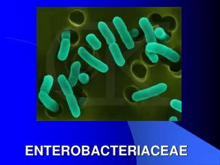
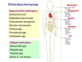
ENTEROBACTERIACEAE • A large Family of aerobic bacterial flora of intestine of humans and other animals. • Its members are nonsporting, non acid-fast, gram negative bacilli. • Capsule ± • Motility ± • General features – aerobic and facultatively anaerobic, grow readily on ordinary media, ferment glucose, reduce nitrates to nitrites and form catalase but not oxidase. .
Wide biochemical and antigenic heterogeneity. • Genetic mechanisms like conjugation and transduction in these bacteria contribute to their infinite variety. • Various classifications of Enterobacteriaceae have been put forward. • Two important classifications are 1. based on taxonomy and 2. based on lactose fermentation. .
CLASSIFICATION BASED ON LACTOSE FERMENTATION • Lactose fermenters • Escherichia coli • Klebsiella sp. • Late lactose fermenters • Shigella sonnei • Para colons etc • No lactose fermenters • Salmonella • Shigella etc. .
Tribe I: Escherichia Genus Escherichia Edwardsville Citrobacter Salmonella Shigella Tribe II: Klebsiella Genus Klebsiella Enterobacter Hafnia Serratia Tribe III: Proteae Genus Proteus Morganella Providencia Tribe IV: Erwinieae Genus Erwinia TAXONOMICAL CLASSIFICATIONENTEROBACTERIACEAE .
Genus Escherichia named after Escherichia who was the first to describe the colon bacillus under the name Bacterium coli commune (1885). • Species: • E.coli, • E.fergusonii, • E.hermanii, • E.vulneris, • E.blattae etc .
MORPHOLOGY • Gram negative bacilli • 1-3 x 0.4-0.7 µm • Single, pairs • Motile by peritrichate flagella • Found in some – capsules, fimbriae, immobility • Non spore forming .
CULTURE CHARACTERISTICS • Aerobe and facultative anaerobe • 10-40°C (37°C) • S = smooth forms seen in fresh isolates, easily emulsifiable in saline. • R = rough forms seen in older cultures, with irregular dull surface, often autoagglutinable in saline. • S-R variation occurs as a result of repeated subcultures and is associated with the loss of surface antigens and usually of virulence. .
Many pathogenic isolates have polysaccharide capsules. • Some strains may occur in the mucoid form. • Nutrient agar – colonies are large, thick, greyish white, moist, smooth, opaque or partially translucent discs. • Blood agar - Many strains esp. pathogenic ones are hemolytic on blood agar. • MacConkey medium - colonies are bright pink due to lactose fermentation. • Broth – general turbidity, heavy deposit. .
BIOCHEMICAL REACTIONS • Sugar fermentation – glucose, lactose, manitol, maltose and many other sugars fermented with acid and gas production. • Sucrose generally not fermented. • IMViC ++-- • Gelatin -, H2S -, urease -. .
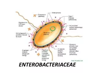
ANTIGENIC STRUCTURE • O = somatic antigen • K = capsular antigen • H = flagellar antigen • So far, >170 types of O, 100 types of H and 75 types of K have been identified. • Antigenic pattern of an organism based on these antigens is written as eg. O111:K58:H2, O54:K27:H41 etc. • K antigen is the acidic polysaccharide antigen located in the envelope or microcapsule (K for kapsel, german for capsule). • It encloses the O antigen and renders the strain inagglutinable by the O antiserum. • It may also contribute to virulence by inhibiting phagocytosis. .
VIRULENCE FACTORS • Surface antigens: O and K • O antigen – somatic lipopolysaccharide surface O antigen has endotoxic activity, protects the bacteria from phagocytosis and bactericidal effects of complement. • K antigen – affords protection against phagocytosis and antibacterial factors in normal serum. .
Fimbriae – plasmid coded, found in small numbers and mediate mannose resistant hem agglutinins, act as virulence factors. • Examples: • CFA = colonization factor antigens in enter toxigenic E.coli causing human diarrhea. • P fimbriae which bind to uroepithelial cells and P blood group substance on human erythrocytes, have a role in urinary tract infection. .
TOXINS: • E.coli produce two kinds of exotoxins – haemolysins and enterotoxins. • Three distinct types of E.coli enterotoxins have been identified – • LT = heat labile toxin • ST = heat stable toxin • VT = serotoxin (also known as SLT = shiga like toxin). .
LT = HEAT LABILE TOXIN • Resembles cholera toxin in its structure, antigenic properties and mode of action. • It is a complex of polypeptide subunits. • Each unit of toxin has 1 subunit A and 5 subunits B. • CT is about 100 times more potent than LT. • LT is a powerful antigen and can be detected by serological and biological tests. .
ST = HEAT STABLE TOXIN • LMW polypeptide, poorly antigenic. • Two types known – STA/ST1 and STB/ST2. • ST genes are carried on plasmids which may also carry other genes, such as for LT and drug resistance. .
VT = VEROTOXIN = VEROCYTOTOXIN • Named so because it was first detected by its cytotoxic effect on Vero cells, a cell line derived from African green monkey kidney cells. • It is also known as SLT = shiga like toxin because it is similar to the shigella dysenteriae type 1 toxin in its physical, antigenic and biological properties. • Has A and B subunits. • VT genes appear to be phage encoded. • VT1, VT2 identified. .
CLINICAL INFECTIONS • URINARY TRACT INFECTIONS • DIARRHEA • PYOGENIC INFECTIONS • SEPTICAEMIA .
URINARY TRACT INFECTIONS • Community acquired: E.coli and other coliforms account for the large majority of naturally acquired UTIs. • Hospital acquired: Those acquired in the hospital, following instrumentation, are more often caused by other bacteria such as pseudomonas and proteus. • The E.coli serotypes commonly responsible for UTI are those normally found in the feces, O groups 1,2,4,6,7,etc. .
Asymptomatic bacteriuria: • Observed in some pregnant women, it means urinary infection without any symptoms. • If it progresses, it may lead to symptomatic infection later in pregnancy, pyelonephritis and hypertension in the pregnant woman, as well as to prematurity and perinatal death of the fetus. • Urinary tract infection can be: • Ascending = via urethra • Descending = haematogenous route .
KASS’ CONCEPT OF SIGNIFICANT BACTERIURIA • Normal urine is sterile, but during voiding may get contaminated by genital commensals. • Hence presence of bacteria in microscopy and culture of a urine sample need not necessarily mean UTI by that organism. • To differentiate between actual pathogen and contaminant, Kass etc. devised the following formula. • Colony count in urine: • <10000/ml = contaminant • 10000 to 100000/ml = indecisive, repeat test. • >100000/ml = significant bacteriuria. • Exceptions: in patients on antibacterial or diuretic drugs and with some bacteria like staph. Aureus, even low counts may be significant. .
URINE SAMPLE COLLECTION AND TRANSPORT • Inform the patient about the procedure. • Proper cleaning of area. • Clean voided midstream urine sample is collected in a sterile leak-proof container. • Sample container is labeled, request form filled and send along with the sample to the laboratory within an hour or two. • If delay of more than 1-2 hours is there, the sample should be refrigerated. • Boric acid can be used as a preservative to prevent the change in count of bacteria in the sample. .
QUANTITATIVE AND SEMIQUANTITATIVE METHODS • Quantitative culture: • Serial ten fold dilutions of urine are tested by the pour plate or surface culture methods. Complicated method. • Semiquantitative culture: • Fixed volume of urine placed on a noninhibitory medium like blood agar with a standard loop for quantitative measurement. • Another loopful of urine placed on a indicator medium like MacConkey agar for presumptive diagnosis of causative organism. • Culture is followed by biochemical tests etc for identification of the pathogen. .
DIARRHEA CAUSING E.COLI • EPEC = ENTEROPATHOGENIC E.COLI • ETEC = ENTEROTOXIGENIC E.COLI • EIEC = ENTEROINVASIVE E.COLI • EHEC = ENTEROHAEMORRHAGIC E.COLI • EAEC = ENTEROAGGREGATIVE E.COLI .
EPEC = ENTEROPATHOGENIC E.COLI • Associated with diarrhea in infants and children. • Institutional outbreaks, sporadic diarrhea. • Do not produce enterotoxins. • Not invasive. • M/A: Attach to mucosa of upper small intestine, cause disruption of brush border microvilli. • Enter adherent E.coli is another name given to them because they can adhere to HEp-2 cells. .
ETEC = ENTEROTOXIGENIC E.COLI • Endemic in developing countries in tropics, all age groups. • Mild watery diarrhea to fatal disease indistinguishable from cholera. • Persons from developed countries visiting endemic areas often suffer from ETEC diarrhea – a condition known as TRAVELER’S DIARRHEA. • Adhere to intestinal epithelium via fimbrial or colonization factor antigens (CFA I,II,III,IV,etc.). • Produce LT or ST or both. • Diagnosis done by demonstration of the toxin. .
EIEC = ENTEROINVASIVE E.COLI • Resemble Shigella • Many are nonmotile, do not ferment lactose or ferment it late with only acid production, and do not form lysine decarboxylase. • Many of these show O antigen cross reactivity with Shigella. • Earlier names given – Shigella alkalescens, Shigella dispar – and were grouped under Alkalescens-Dispar group. • Named EIEC because they have the capacity to invade interstitial epithelial cells in vivo and penetrate HeLa cells in tissue culture. .
Clinically EIEC infection resembles shigellosis, ranging from mild diarrhea to frank dysentery. • Sereny test: • Instillation of a suspension of freshly isolated EIEC or Shigella into the eyes of guinea pigs leads to mucopurulent conjunctivitis and severe keratitis. • Mice can also be used. • Cell Penetration in HeLa or HEP-2 cells. • Plasmid detection: • VMA ELISA: The plasmid codes for outer membrane antigens called the virulence marker antigens (VMA) which can be detected by the ELISA (VMA ELISA) test. .
EHEC = ENTEROHAEMORRHAGIC E.COLI • Produce VT • Mild diarrhea to fatal hemorrhagic colitis and hemorrhagic uremic syndrome (HUS) particularly in young children and elderly. • Primary target of VT = vascular endothelial cells. • O157:H7, O26:H1 etc • The disease may occur sporadically or as outbreaks of food poisoning. • Changing lifestyle and eating habits. • Salad vegetables such as radish and alfalfa sprouts, in which bacteria were found beneath the skin and in the deeper tissues. • Diagnosis: demonstration of VT. .
EAEC = ENTEROAGGREGATIVE E.COLI • Appear aggregated in a stacked brick formation on Hep-2 cells or glass. • They have been associated with persistent diarrhea, especially in developing countries. • They form a LMW heat stable enterotoxin called EAST1 (enter aggregative heat stable enterotoxin-1). .
PYOGENIC INFECTIONS: • E.coli form the most common cause of intra-abdominal infections, such as peritonitis and abscess resulting from spillage of bowel contents. • Pyogenic infections in the perianal area. • Neonatal meningitis • SEPTICAEMIA: • Blood stream invasion by E.coli may lead to fatal conditions like septic shock and systemic inflammatory response syndrome (SIDS). • HeLa = Human carcinoma of cervix cell line (named after a lady named Hela) • HEP-2 = Human epithelioma of larynx cell line • Vero = vervet monkey kidney cell line .