Download
1 / 31
330 likes | 503 Views
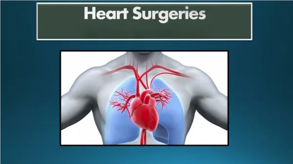
3D ANATOMICAL BASIS FOR TRANSOBTURATOR SURGERIES. Prof. Paulo Palma. Paradigm shifts. 1. Integral Theory, 1993 (Peter Petros) Creating neoligaments using tapes Reinforcement of pubourethral ligament Midurethral tape placement. 2. Transobturator Approach, 2001 (Delorme)

E N D
3D ANATOMICAL BASIS FOR TRANSOBTURATOR SURGERIES Prof. Paulo Palma
Paradigm shifts 1. Integral Theory, 1993 (Peter Petros) • Creating neoligaments using tapes • Reinforcement of pubourethral ligament • Midurethral tape placement. 2. Transobturator Approach, 2001 (Delorme) - Reinforcement of urethropelvic ligament - Less visceral and vascular complications - Less irritative voiding symptoms
Urethropelvic Ligament Compartiments Anterior Median Posterior Uterus Sacrum P Bladder Pubourethral Ligament Sacro Uterine Ligament Tendineous Arc Vagina
LIGAMENTS Sacrum P Pubourethral Ligament Sacro Uterine Ligament Tendineous Arc Vagina Urethropelvic Ligament
First muscular layer 1. Superficial Transverse Muscle of perineum 2. Isquiocavernosous muscle 3. Bulbocavernosous Muscle 4. External Anal Esfincter 3 2 1 4
Second Muscular Layer 5 5.Deep Transversal Muscle of perineum
White Line 6 6. Pubovaginal 7. Puborectal 8. Ileococcigeous 7 8
Bony Anatomy Obturator Canal Pubic Symphysis Ischial tuberosity Ischiopubic ramus
Muscular Anatomy Gracilis Adductor longus Clitoris Adductor magnus
Obturator VesselsRetroperitoneal View Obturator nerve Obturator vessels Obturator canal
Obturator - Neuroanatomy • From anterior division of L2-4 • Innervates all muscles of the obturator region, except pectineus • Passes through obturator canal and splits to innervate the muscles that adduct the thigh and help with external rotation • Small sensory area on the medial thigh
Anterior aginal Wall Defects Uterus Tendineous Arc P Vagina
Lateral Defect Anterior aginal Wall Defects Uterus Tendineous Arc P Vagina
Anterior aginal Wall Defects LateralDefect
Central Defect Central Defect Uterus Tendineous Arc P Vagina
Anterior Vaginal Prolapse (AVP) Uterus Tendineous Arc P Pubourethral Ligament Vagina
Anterior Vaginal Prolapse (AVP) Central Defect Uterus P Tendineous Arc Lateral Defect Vagina
Anterior Vaginal Prolapse (AVP) Uterus P Vagina
Evaluation of pelvic floor reconstructive surgery using tridimentional helical CT Fig 3. Notice the well supported bladder base and the TOT arms
Posterior Defect Posterior Vaginal Prolapse (PVP) Rectum Uterus Tendineous Arc P Vagina Levator ani
Posterior Vaginal Prolapse Repair Rectum Uterus Tendineous Arc P Sacrospinal Ligament Vagina Levator ani
Evaluation of pelvic floor reconstructive surgery using tridimentional helical CT Fig 4. Infracoccigeal sacropexy showing indirectly the “neo rectovagianl fascia”