Download
1 / 71
810 likes | 1.38k Views
Prevention of Diabetic Foot Ulcers and Lower Extremity Amputation. Barry Stults, MD Scott A. Clark, DPM Thomas Miller, MD. © 2007. American College of Physicians. All rights reserved. This content has been excerpted from the ACP Clinical Skills Module, "Diabetic Foot Ulcers."

E N D
Prevention ofDiabetic Foot Ulcers and Lower Extremity Amputation Barry Stults, MD Scott A. Clark, DPM Thomas Miller, MD © 2007. American College of Physicians. All rights reserved. This content has been excerpted from the ACP Clinical Skills Module, "Diabetic Foot Ulcers." For more information visit: http://www.acponline.org/clinicalskills/
“…the enormity of the global burden of diabetic foot disease…this much neglected, but potentially devastating, complication of a disease that is reaching epidemic proportions…Someone, somewhere, loses a leg because of diabetes every 30 seconds of everyday…” Lancet. 2005;366:1674
Case Study • 64-year-old obese man • Type 2 DM (15 yrs) • BP (18 yrs) • Dyslipidemia (18 yrs) • CABG (10 yrs ago) • Claudication (today; 25 yds) • Insulin/Metformin/Statin/ACEI/HCTZ/ASA • “Sore on my left foot, Doc”
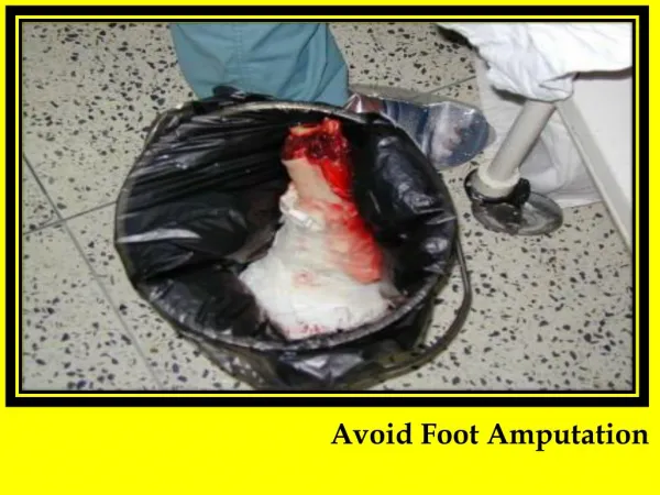
Case Study (continued) • Clinical evaluation of heel ulcer: • Probe reached bone • Extensive subcutaneous abscess • MRI: extensive osteomyelitis • ABI: 0.2 • Angiography: Inoperable severe vascular disease • Uncontrolled infection • Amputation necessary
Amputations in Diabetes Common: • U.S.A. – 80,000 amputations/year (2002) Costly: • $60,000/amputation • $2 billion total costs annually Lancet. 2005;366:1719 DiabetesCare. 2004;27:1598 DiabetesCare. 2003;26:495
Tragic “Rule of 50” Transfemoral/transtibial level 2nd amputation in 5 years Die in 5 years • 50% ofamputations • 50% of patients • 50% of patients ClinicalCareoftheDiabeticFoot, 2005
Tragic “Rule of 15” • 15% of diabetes Foot ulcer in lifetimepatients • 15% of foot ulcers Osteomyelitis • 15% of foot ulcers Amputation ClinicalCareoftheDiabeticFoot, 2005
Team Care • Identification of high-risk patients • Detection of early problems • Educate/motivate self-care behaviors • Prophylactic nail/skin care • Therapeutic footwear • Prompt, multidisciplinary treatment of ulcers Lancet. 2005;366:1676
Team Care Reduces Ulcers/Amputations • 50%-80% reductions in ulcers/amputations • Economic modeling studies • Cost-effective if 25%-40% reduction in ulcer rate • Cost-saving if > 40% reduction in ulcer rate Lancet. 2005;366:1719 DiabetesCare. 2004;27:901
% Causal Pathways Neuropathy: 78% Minor trauma: 79% Deformity: 63% Behavioral ? Neuropathy Causal Pathways for Foot Ulcers Deformity Minor Trauma - Mechanical (shoes) - Thermal - Chemical Poor self-foot care ULCER DiabetesCare. 1999; 22:157
Detecting Feet-at-risk • History: • Prior amputation or foot ulcer • Peripheral artery disease (PAD) • Exam: • Insensate • Foot deformities • Absent pulses • Prolonged venous filling time • Reduced ABI • Pre-ulcerative cutaneous pathology ArchInternMed. 1998;158:157
Risk Stratify for Ulcer Risk DiabetesCare. 2001;24:1442 DiabetesMetab. 2003;29:261
Annual Diabetic Foot Exams 2000 Behavioral Risk Factor Surveillance System, CDC *p < 0.01 HealthServicesResearch. 2005;40:361
Sensory Neuropathy in Diabetes • Loss of protective sensation in feet • Detect with 5.07/10-g Semmes-Weinstein monofilament • 50% of insensate patients have no symptoms DiabetesCare. 2006;29(Suppl 1):S24 DiabetesCare. 2004;27:1591
Monofilament Testing • Test characteristics: • Negative predictive value = 90%-98% • Positive predictive value = 18%-36% • Prospective observational study: • 80% of ulcers and 100% of amputations occur in insensate feet • Superior predictive value vs. other test modalities • JFamPract. 2000;49:S30 • DiabetesCare. 1992;15:1386
Using the Monofilament • Demonstrate on forearm or hand • Place monofilament perpendicular to test site • Bow into C-shape for 1 second • Test 4 sites/foot • Heel testing does notpredict ulcer • Avoid calluses, scars, and ulcers
Monofilament Testing Tips • Insensate at 1 site = insensate feet • Falsely insensate with edema, cold feet • Test annually when sensation normal • Use monofilament • < 100 times day • Replace if bent • Replace every 3 months
Vibration Testing • Biothesiometer • Best predictor of foot ulcer risk • 128-Hz tuning fork at halluces • Equivalent to 10-g monofilament • Newly recommended by ADA DiabetesCare. 2006;29(Suppl 1):S25 DiabetesResClinPract. 2005;70:8
Motor Neuropathy and Foot Deformities • Hammer toes • Claw toes • Prominent metatarsal heads • Hallux valgus • Collapsed plantar arch
Hammer Toes Claw Toes © 2002 American Diabetes Association From The Uncomplicated Guide to Diabetes Complications Reprinted with permission from The American Diabetes Association
Hallux Valgus © 2002 American Diabetes Association From The Uncomplicated Guide to Diabetes Complications Reprinted with permission from The American Diabetes Association
Boulton, et al. Guidelines for Diagnosis of Outpatient Management of Diabetic Peripheral Neuropathy. DiabeticMedicine 1998, 15:508-512
Pre-ulcer Cutaneous Pathology • Persistent erythema after shoe removal • Callus • Callus with subcutaneous hemorrhage • Fissure • Interdigital maceration, fungal infection • Nail pathology
Pre-ulcer AJM Boulton, H Connor, PR Cavanagh, The Foot in Diabetes, 2002
Peripheral Artery Disease • Prevalence (ABI < 0.9): • 10%-20% in type 2 diabetes at diagnosis • 30% in diabetics age 50 years • 40%-60% in diabetics with foot ulcer • Complications: • Claudication • Associated coronary and cerebral vascular disease • Delayed ulcer healing DiabetMed. 2005;22:1310 DiabetesCare. 2003;26:3333
Pedal Pulse Examination • Absent pedal pulses predicts severe PAD • Absence of a single pedal pulse does not predict PAD • Presence of pedal pulses does not rule out PAD! • ArchInternMed. 1998;158:1357 • DiabetesCare. 2003;26:3333
Venous Filling Time • Sitting: Locate pedal vein bulging above skin • Supine: Elevate leg to 45° for 1 minute • Sitting: Check time to pedal vein bulging • JClinEpidemiol. 1997;50:659 • ArchInternMed. 1998;158:1357
Venous Filling Time Interpretation Filling Time Normal <20 sec Abnormal/collaterals 20-40 sec Severe PAD >40 sec • Filling time > 20 sec predicts ABI < 0.5 • Sensitivity, 22%; Specificity, 94%; LR, 3.9 • JClinEpidemiol. 1997;50:659 • ArchInternMed. 1998;158:1357
Adapted from: Norman PE, Eikelboom JW, Hankey GJ. Peripheral arterial disease: prognostic significance and prevention of atherothrombotic complications. MedicalJournalofAustralia 2004; 181:150-154. Figure 1, p.151
Ankle-Brachial Index • Screening: 2004 ADA recommendation • “Consider” at age 50 years and every 5 years • Diagnosis: • Claudication, absent DP/PT pulses, foot ulcer • Limitations: • Underestimates severity in calcified arteries DiabetesCare. 2005;28:2206 DiabetesCare. 2004;27(Suppl 1):S15-S35