Download

1 / 22
230 likes | 652 Views
Brightfield and Phase Contrast Microscopy. Real and virtual image formation by biconvex lenses. Lens focal point For an object further away than the lens focal point, an inverted, real image will be formed on the opposite side of the lens

E N D
Real and virtual image formation by biconvex lenses • Lens focal point • For an object further away than the lens focal point, an inverted, real image will be formed on the opposite side of the lens • For an object closer than the focal point, a virtual image will be formed on the same side of the lens • http://micro.magnet.fsu.edu/primer/java/lens/bi-convex.html
Compound Microscope • The compound microscope uses at least two lens systems • The objective forms an intermediate real image of the object at the objective tube length • The ocular forms a virtual image of that intermediate image to the retina of the eye • If we are dealing with a photodetector, we must use a projection lens to form a real image from the intermediate image
Current microscope objective tend to be infinity corrected • Infinite tube length • Require an additional lens in objective to converge beam • Advantages • Objectives are simpler • Optical path is parallel through the microscope body:
Other lenses • Collecter • Condenser • Allow us to use point light sources instead of parallel illumination • Also (later) increase the resolution of the microscope • Ironically, van Leeuwenhoek, who used simple non-compound, single-lens microscopes, was using the lens of his eye as a projection lens!
Lens Resolution • Geometric optics predicts lenses of infinite resolution • However, because of the phenomenon of diffraction, every point in the object is converted into an Airy disc • Diameter of Airy disc: D = 1.22 X λ / n sin α, or D = 1.22 X λ / NA
We cannot resolve objects whose Airy discs overlap by ~20% As a consequence, Abbe’s rule is that d=λ/NA http://micro.magnet.fsu.edu/primer/java/microscopy/airydiscs/index.html
Reading an objective http://micro.magnet.fsu.edu/primer/anatomy/specifications.html
For a typical 1.3 NA lens at 525 nm, the limit of resolution is ~ 400 nm • How to improve? • Larger NA (lenses, immersion fluid) • Shorter λ • Add a condensor: D = λ / (NAobj. + NAcond.) • So, for a 1.3 NA lens and condensor, D drops to ~200 nm
Abberations • Spherical aberration • Most severe • Immersion fluid • Field curvature • Chromatic aberration • Astigmatism, coma • http://micro.magnet.fsu.edu/primer/lightandcolor/opticalaberrations.html
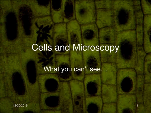
Brightfield microscopy • Generally only useful for stained biological specimens • Unstained cells are virtually invisible
H&E stain From Wikipedia, the free encyclopedia H&E stained lung tissue sample from an end-stage emphysema patient. RBCs are red, nuclei are blue-purple, and other cellular and extracellular material is pink. H&E stain, or haematoxylin and eosin stain, is a popular staining method in histology. It is the most widely used stain in medical diagnosis; for example when a pathologist looks at a biopsy of a suspected cancer, the histological section is likely to be stained with H&E and termed H&E section, H+E section, or HE section. The staining method involves application of the basic dye haematoxylin, which colors basophilic structures with blue-purple hue, and alcohol-based acidic eosin Y, which colors eosinophilic structures bright pink. The basophilic structures are usually the ones containing nucleic acids, such as the ribosomes and the chromatin-rich cell nucleus, and the cytoplasmatic regions rich in RNA. The eosinophilic structures are generally composed of intracellular or extracellular protein. The Lewy bodies and Mallory bodies are examples of eosinophilic structures. Most of the cytoplasm is eosinophilic. Red blood cells are stained intensely red. The structures do not have to be acidic or basic to be called basophilic and eosinophilic. The terminology is based on the affinity to the dyes. Other colors, e.g. yellow and brown, can be present in the sample; they are caused by intrinsic pigments, e.g. melanin. Some structures do not stain well. Basal laminae need to be stained by PAS stain or some silver stains, if they have to be well visible. Reticular fibers also require silver stain. Hydrophobic structures also tend to remain clear; these are usually rich in fats, eg. adipocytes, myelin around neuronaxons, and Golgi apparatus membranes.
Radiolarian in Darkfield http://micro.magnet.fsu.edu/primer/techniques/darkfield.html
Phase contrast http://microscopy.fsu.edu/primer/techniques/phasegallery/chocells.html