Download
1 / 58
620 likes | 883 Views
DNA structure- Part II. Levels of DNA structure. Levels of DNA structure. 1°structure: the order of bases on the polynucleotide sequence; the order of bases specifies the genetic code. 2°structure: the three-dimensional conformation of the polynucleotide backbone.

E N D
DNA structure- Part II Levels of DNA structure
Levels of DNA structure • 1°structure: the order of bases on the polynucleotide sequence; the order of bases specifies the genetic code. • 2°structure: thethree-dimensional conformation of the polynucleotide backbone. • 3°structure: supercoiling. • 4°structure: interaction between DNA and proteins
DNA - 1° Structure • Deoxyribonucleic acids (DNA) is a biopolymer that consists of a backbone of alternating units of 2-deoxy-D-ribose and phosphate.
Continue…. This bond which indicates that there are two covalent bonds formed between the -OH and the acidic phosphate group • The 3’-OH of one 2-deoxy-D-ribose is joined to the 5’-OH of the next 2-deoxy-D-ribose with bases by a phosphodiester bond.
Continue…. • So, the Primary Structureis the sequence of bases along the pentose-phosphodiester backbone of a DNA molecule • Base sequence is read from the 5’ end to the 3’ end. • System of notation single letter (A,G,C,U and T).
The backbone of DNA & RNA is hydrophilic. • The hydroxyl gp (OH) of the sugar residues form hydrogen bonds with water. • The Phosphate gp, with pka 2, are completely ionized and –vely charged at pH 7. • The –ve charges are neutralized by ionic interactions with +ve charges on proteins, metal ions. • A short nucleic acid containing 50 or fewer nucleotides is called an oligonucleotide, a longer is a polynucleotide.
the 2’ hydroxyl is absent in DNA
Abbreviations of Nucleic Acid Sequences • pA-C-G-T-AOH • or pApCpGpTpA • or pACGTA
Nucleotide Bases affect the three dimensional structure of nucleic acids • The purine and pyrmidine bases are conjugated ring system. • One result is that pyrmidines are planar molecules and purines are nearly planar with a slight pucker. • Free purine and pyrimidine bases can exist in two or more forms called tautomers, depending on pH. A tautomeric shift occurs when a proton changes its position, resulting in a rare tautomeric form.
Continue…. • Standard and anomalous base-pairing arrangements that occur if bases are in the rare tautomeric forms. Base mispairings due to tautomeric shifts were originally thought to be a major source of errors in replication. • Such structures have not been detected in DNA, and most evidence now suggests that other types of anomalous pairings are responsible for replication errors.
Continue…. • As a result of resonance, delocalized electrons in the conjugated ring rings are available to absorb UV light at 260nm. • The chemical properties of the purine and pyrimidines give rise to two modes of interaction between bases: • Hydrophobic stacking, the bases relatively insoluble in water at neutral pH. • Base pairing which result from H-bonding . As a result of resonance, all nucleotide bases absorb UV light.
Nucleotides play additional roles in cells • Adenosine is a building block for some important enzyme cofactor, such as NAD+ and FAD. The presence of an adenosine component in a variety of cofactors enables recognition by enzymes that share common structural features. NAD+ FAD
Continue…. • cAMP formed from ATP in a reaction catalyzed by adenylyl cyclase, is a common second messenger produced in response to hormones and other chemical signals
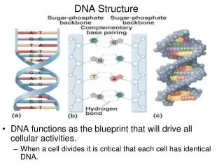
DNA - 2° Structure • Secondary structure is the ordered arrangement of nucleic acid strands. • Double helix is a type of 2° structure of DNA molecules in which two anti-parallelpolynucleotide strands are coiled in a right-handedmanner about the same axis. • Structure based on X-Ray crystallography. • The molecule is stabilized by hydrophobic interactions in its interior and by hydrogen bonding between the complementary bases pairs A-T and G-C
Base Pairing • Base pairing is complementary. • A major factor stabilizing the double helix is base pairing by hydrogen bondingbetween T-A and between C-G • T-A base pair comprised of 2 hydrogen bonds. • G-C base pair comprised of 3 hydrogen bonds
Base stacking • Bases are hydrophobic and interact by van der Waals interactions. • In standard B-DNA, each base rotated by 32° compared to the next and, while this is perfect for maximum base pairing, it is not optimal for maximum overlap of bases; in addition, bases exposed to the minor groove come in contact with water. • Many bases adopt a propeller-twistin which base pairing distances are less optimal but base stacking is more optimal and water is eliminated from minor groove contacts. • The propeller twist of the base pairs results in purine-purine clash in the center of the helix. Because the purines are larger than the pyrimidine rings, they extend beyond the helical axis of DNA. Hydrophobic, van der Waals, and electrostatic interactions favor the alignment of bases in an aqueous solution or within a polynucleotide chain the un-stacked orientation is disfavored.
DNA attempts to reduce purine-purine clash in several ways: A. The base pairs rotate less along the helix axis in the purine-pyrimidine sequences (lower average helical twist). They tend to rotate less in the pyrimidine-purine sequences (lower than average helical twist). The average helical twist was still very close to the 36° proposed by Watson and Crick. • Another way DNA minimizes the purine-purine clash is that it bends toward the minor grove or major groove to reduce the interaction. C. Finally clashing base pairs could slide left or right toward the phosphodiester backbones to minimize the purine-purine interaction.
Major and minor grooves • The major groove is large enough to accommodate an alpha helix from a protein. • Regulatory proteins (transcription factors) can recognize the pattern of bases and H-bonding possibilities in the major groove.
DNA adopts different helical forms • Nucleic acids are inherently flexible molecules. Numerous bonds in the sugar-phosphate backbone can rotate, and thermal fluctuation can lead to bending, stretching, and un-pairing of the two strands. • As a result, cellular DNA contains significant deviations from the Watson-Crick DNA structure, some or all of which may play important roles in DNA metabolism. • Generally, such structural variations do not affect the key properties of strand complementarity: antiparallel strands and the requirement for AT and G≡C base pairs.
Variation in the three-dimensional structure of DNA reflect three things: (1) The different possible confirmations of the deoxyribose. (2) Rotation about the contiguous bonds that make up the sugar-phosphate backbone. (3) Free rotation about the glycosidic bond.
Continue…. • B-DNA (Watson-Crick Form) • considered the physiological form • aright-handed helix, diameter 11Å • 10 base pairs per turn (34Å) of the helix. • A-DNA (favored in environment with very • low water content) • aright-handedhelix, but thicker than B-DNA. • 11 base pairs per turn of the helix • has not been found in vivo. • Z-DNA occurs at high salt concentrations in • polymers that have a sequence of alternating purines and pyrimidines • aleft-handeddouble helix. • may play a role in gene expression. • Z-DNA occurs in nature, usually consists of alternating purine-pyrimidine bases. • Methylated cytosine found also in Z-DNA.
In each case, the sugar-phosphate backbones wind around the exterior of the helix (red and blue), with the bases pointing inward. The same 25-base-pair DNA sequence is shown in all three forms. Differences in helical diameter can be seen in end-on views (top); differences in helical rise and groove shape are apparent in the side views (bottom). B-DNA, the most common form in cells, has a wide major groove and a narrow minor groove. A-form helices, common for RNA and certain DNA structures, are more compact than B-DNA. The major groove is deeper and the minor groove is shallower than in B-DNA. Z-DNA, which forms only under high salt conditions or with C≡G-rich DNA sequences, is left-handed, and its backbone has a zigzag pattern. It is less compact than B-DNA, with a very shallow major groove and a narrow and deep minor groove.
DNA - 3° Structure • Tertiary structure is the three-dimensional arrangement of all atoms of a nucleic acid; commonly referred to assupercoiling. • The term "supercoiling" means literally the coiling of a coil. • DNA is coiled in the form of a double helix. • Enzymes called topoisomerases or gyrases can introduce or remove supercoils. • If there is no net bending of the DNA axis upon itself, the DNA is said to be in a relaxed state. • Negative supercoiling may promote cruciforms
Certain DNA Sequences Adopt Unusual Structures • Other sequence-specific DNA structures have been detected, within larger chromosomes, that may affect the function and metabolism of the DNA segments in their immediate vicinity. • For example, certain repetitive sequences can bend the DNA helix in a distinct way. • This DNA bending helps certain proteins—such as transcription factors, which promote the synthesis of mRNAs—bind to their target DNA binding sites. • Regions of DNA where the two complementary strands have the same sequence when read in the 5′→3′ or the 3′→5′ direction occur relatively frequently in chromosomal DNA and are called palindromes.
Continue…. • In language, a palindrome is a word, phrase, or sentence that is spelled identically when read either forward or backward. • Two examples are ROTATOR and NURSES RUN. • In biology, the term applies to double-stranded regions of DNA where one strand’s sequence is identical to its complement. • for example, 5′-GAATTC-3′ is a palindrome because its complementary sequence is also 5′-GAATTC-3′. • Palindromes are formed from adjacent inverted repeats, which can occur within one strand of DNA or over the two strands of the double helix
Continue…. • These sequences play important biological roles, such as: • Slowing or blocking protein synthesis by the ribosome-a process called translation attenuation . • Forming recognition sites for restriction enzymes, which catalyze double-stranded DNA cleavage.
DNA classes and sizes Circular DNA is a type of double-stranded DNA in which the 5’ and 3’ ends of each stand are joined by phosphodiester bonds. Human chromosome DNA Plasmid DNA E. Coli DNA λ phage DNA
Gene What is in the middle?????? Something Happens Protein
RNA Structure • In the early 1970s, Alexander Rich, Aaron Klug, and Sung-Hou Kim independently solved the structures of transfer RNAs, revealing how tRNAs carry the amino acids that are used in protein synthesis on the ribosomes. • The wide-ranging functions of RNA reflect a structural diversity much richer than that observed in DNA molecules.
Continue…. • The single strand of RNA folds back on itself to form short base-paired or partially base-paired segments connected by unpaired regions. • This property, called RNA secondary structure, enables RNA molecules to fold into many different shapes that lend themselves to many different biological functions.
Continue…. • The greater structural variety in RNA relative to DNA reflects the three main chemical differences between the two polynucleotides: 1- The pentose (2′-deoxyribose in DNA vs. ribose in RNA). 2-The base composition (thymidine vs. uridine). 3-The sugar pucker of the pentose (C-2′ endo vs. C-3′ endo). • Double-stranded RNAs do exist in nature, such as those that form the genomes of some viruses. • In addition, some RNAs do not seem to form stable three-dimensional structures from local base-pairing interactions, e.g. mRNA. • These RNAs may fold into three-dimensional structures only in the presence of bound proteins, forming complexes called ribonucleoproteins (RNPs).
RNAs Form Various Stable Three-Dimensional Structures • Most of the highly structured RNAs contain noncanonical base pairs and backbone conformations not observed in DNA. • In many cases, the 2′-hydroxyl group on ribose, a chemical feature that distinguishes RNA from DNA, seems to be directly or indirectly responsible for these unique structural properties. • The presence of the 2′ hydroxyl makes RNA vulnerable to hydrolysis, but it also allows for additional hydrogen bonding between segments of an RNA molecule. • As a result, RNA helices are more thermodynamically stable than are DNA helices of the same length and sequence.
Continue…. • Base pairs other than canonical AU and CG pairs are common in RNAs, including A-A and G-U. In all cases, base pairs or single bases are most stable when stacked on top of one another in a helix. • Divalent and monovalent metal ions (Mg2+, Ca2+, K+, and Na+) bind to specific sites in RNA and help shield the negative charge of the backbone, allowing parts of the molecule to pack more tightly together.
RNA is very similar to DNA Chemically, RNA is very similar to DNA. There are some main differences: 1- RNA uses the sugar ribose instead of deoxyribose in its backbone. Vicinal Hydroxyl Group Makes Difference !! • The vicinal OH groups of RNA are susceptible to nucleophilic attach leading to hydrolysis of the phosphodiester bond. • DNA is not susceptible to alkaline hydrolysis. RNA is alkali labile.
Continue… 2-RNA uses the base Uracil (U) instead of Thymine (T). U is also complementary to A. But Why DNA Contains Thymine rather than Uracil?! • It is because cytosine deaminates to form uracil in a finite rate in vivo. This would results in a mutation in the DNA: • So any U found in DNA will be corrected by a “proofreads” system. Thus DNA can not normally have U. Guanine and cytosine form a base pair stabilised by three hydrogen bonds, whereas adenine and thymine bind to each other through two hydrogen bonds. The red frames highlight the functional groups of cytosine and thymine that are responsible for forming the hydrogen bonds. Cytosine can spontaneously undergo hydrolytic deamination, resulting in a uracil base with the same capability for hydrogen bond formation as thymine.
Continue… 3- RNA tends to be single-stranded. 4-Functional differences between RNA and DNA. • DNA single function, RNA many functions according to their type. • Example of types of RNA e.g: tRNA, mRNA, rRNA.
The Central Dogma • The Flow of Information: DNA → RNA → Protein. • A gene is expressed in two steps: • DNA is transcribed to RNA. • Then RNA is translated into protein.
Many RNA molecules do not encode proteins • tRNA. • rRNA. • a vast number of other non-coding RNAs (ncRNAs). - 98% of the human genome does not code for protein. What is its function?
How big part of human transcribed RNAresults in proteins? • Of all RNA, transcribed in higher eukaryotes, 98% are never translated into proteins. • Of those 98%, about 50-70% are introns. • 4% of total RNA is made of coding RNA. • The rest originate from non-protein genes, including rRNA, tRNA and a vast number of other non-coding RNAs (ncRNAs). • Even introns have been shown to contain ncRNAs, for example snRNAs. • It is thought that there might be order of 10,000 different ncRNAs in mammalian genome
The “Other” 98% of the Human Genome • ncRNA genes have diverse and essential roles. • May be relics of ancient RNA-based life. • Many cellular machines contain RNA. • Ribosome rRNA • Spliceosome snRNAs (U1,U2,U4,U5,U6) • Telomerase Telomerase RNA
RNA types mRNA Non-coding RNA. Transcribed RNA with a structural, functional or catalytic role rRNA Participate in protein synthesis snRNA found in nucleolus, involved in modification of rRNA. tRNA Interface between mRNA & amino acids snRNA: RNA that form part of the spliceosome miRNA involved regulation of expression Other large RNA with roles in chromotin structure stRNA- Small temporal RNA, with a role in developmental timing siRNA- Small interfering RNA, Active molecules in RNA interference
RNA molecules are classified according to their structure and function
tRNA • The smallest kind of the three RNAs. • A single-stranded polynucleotide chain between 73-94 nucleotide residues. • Intramolecular hydrogen bonding occurs in tRNA. • tRNA is a cloverleaf shaped, single strand. • It has: • Anticodon loop. • Amino acid binding site at the 3’end. • Two other loops for binding the ribosomes.
rRNA • rRNA: a ribonucleic acid found in ribosomes, the site of protein synthesis. • Only a few types of rRNA exist in cells. • Ribosomes consist of 60 to 65% rRNA and 35 to 40% protein. • In both prokaryotes and eukaryotes, ribosomes consist of two subunits, one larger than the other.
mRNA • A ribonucleic acid that carries coded genetic information from DNA to ribosomes for the synthesis of proteins. • Present in cells in relatively small amounts and has a short-half life. • Single stranded and unstable. • Biosynthesis is directed by information encoded on DNA. • A complementary strand of mRNA is synthesized along one strand of an unwound DNA, starting from the 3’ end.
Eukaryotic mRNA Structure • Capping: linkage of 7-methylguanosine 7 to the 5’ terminal residue. • Tailing: attachment of an adenylate polymer (poly A)
Chemical and thermodynamic importance of the DNA structure Denaturation and renaturation Complementary base paring by hydrogen bonding and van der Waals interaction → storage and transfer of genetic information
Properties of DNA- Denaturation • When DNA is heated to 80OC, its UV absorbance increases by 30-40% . • With heating, noncovalent forces holding DNA strands together weaken and break. • When the forces break, the two strands come apart in denaturation or melting.
Denaturation of DNA • As strands separate, absorbance at 260 nm increases. • Stacked base pairs in native DNA absorb less light . • Increase is called hyperchromicity. • Temperature at which DNA strands are ½ denatured is the melting temperature or Tm. • Melting Temperature (Tm) – the temp. at which half of the helical structure is lost - What is the hyperchromic shift Hyperchromic Effect – a large increase in light absorbance at 260 nm occurring as double-helical DNA is melted (i.e. unwound).