Download
1 / 32
320 likes | 339 Views
Explore the intricate process of the cell cycle, focusing on cell division, mitosis, and key phases like prophase, metaphase, anaphase, and telophase. Learn about the role of chromosomes, DNA duplication, and the significance of cytokinesis.

E N D
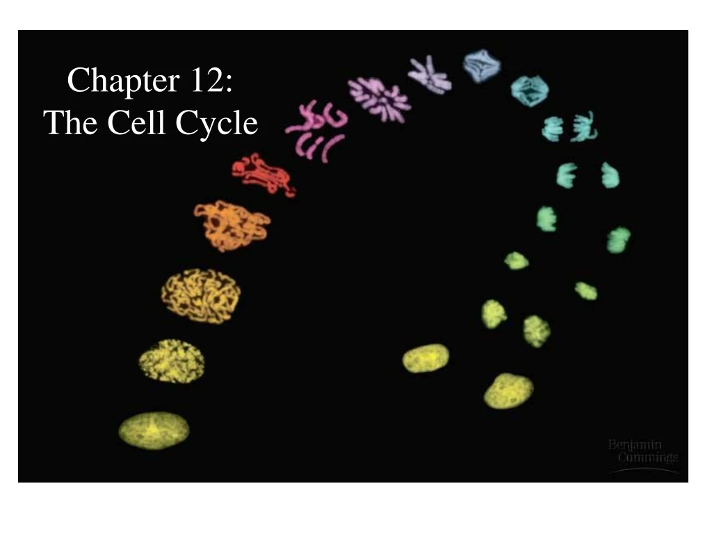
Chapter 12: The Cell Cycle
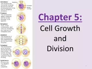
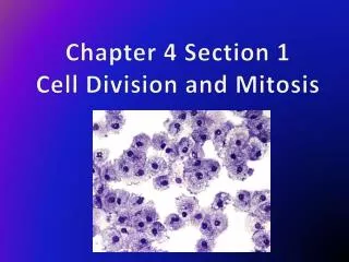
The Cell Cycle A. The Role of Cell Division Purposes of Cell Division (Or, one reason we need that ATP from Cellular Respiration (Chapter 9): a. Single celled organisms (e.g. the Protista) can replicate by reproducing the entire organism, e.g. an amoeba; the process is called mitosis b. Allows reproduction for multicellular organisms by producing female and male gametes (egg, sperm) in the reproductive tissues; the process is called meiosis (Meiosis is described in Chapter 13) c. Growth and also replacement of aged cells for multicellular organisms – these are the somatic cells and the process is also called mitosis and is described here in Chapter 12
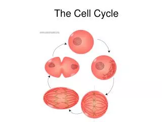
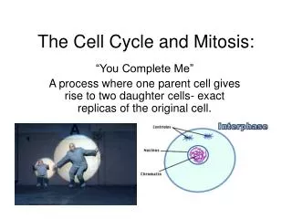
- Process of mitosis: • The aim is to produce two identical daughter cells each containing exact replicas of the mother cell’s chromosomes (this means the entire genome = all genes need to be duplicated exactly). • Thus, all the DNA must be copied so there are two complete sets, one set for each daughter cell. • The outline of the replication process for a chromosome is given in Figure 12.4 (p. 220): Chromosome duplication and distribution during mitosis. • Remember the terms used in this Figure!
Note that: - Genes are present in structures called chromosomes - Each eukaryotic cell has a nucleus (Chapter 6) containing a species-specific number of chromosomes. The human nucleus contains 46 chromosomes 23 from egg, 23 from sperm that fertilize the egg. This means that there are two of each type of chromosome in our cells and there are at least two of each type of gene. There will be an exception to this that we will see later (sex-related genes). - Each chromosome contains hundreds or thousands of genes (these make up our inherited traits). - The DNA that makes up the chromosomes is complexed with proteins this is called chromatin. The proteins maintain the shape of the chromosome.
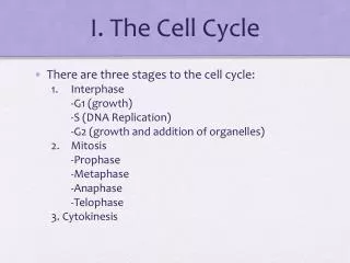
B. Mitotic Cell Cycle exists in four phases: 1. G1 Phase 1st growth phase 2. S Phase DNA duplicated 3. G2 Phase Final growth phase 4. Mitosis/Cytokinesis Figure 12.5 (p. 221) – Note that G1, S, and G2 phases are together called interphase and represent 90% of the cells growth cycle. During interphase, the cell has not started to divide into two daughter cells, but is growing and preparing for division. This is illustrated later in a short movie. Division takes place during the mitotic phase (10% of the cell’s growth cycle). In this phase, mitosis divides the nucleus into two nuclei and cytokinesis (cell division) divides the cytoplasm into two cells.
Mitosis itself can be divided into distinct phases as follows (Note, this all needs to be memorized): • 1. Prophase • 2. Prometaphase • 3. Metaphase • 4. Anaphase • 5. Telophase • You need to pay attention and memorize Figure 12.5 and the information that it contains!
Prophase • - Nucleolus disappears • - Chromatin condenses, this causes the chromosomes to begin to become visible as discrete units • - Centrosomes separate, moving to opposite ends of the cell. • - The centrosomes start to form a framework used to separate the two sister chromatids called the mitotic spindle, that is made of microtubules
2. Prometaphase - Nuclear envelope fragments - Chromosomes become more condensed - A kinetochore is formed at the centromere, the point where the sister chromatids are attached - Microtubules attach at the kinetochores, leading back to the spindle poles (where the centrioles now reside
3. Metaphase - Chromosomes align on an axis called the metaphaseplate. This is a very distinct aspect of metaphase. - Note: the spindle consists of microtubules, one attached to each chromosome Figure 12.7 (p. 224) – The mitotic spindle at metaphase.
4. Anaphase - Paired centromeres separate - One chromatid goes toward each pole - Cell begins to elongate, caused by microtubules not associated with the kinetochore
5. Telophase - Formation of nuclear membrane and nucleolus - Formation of the cleavagefurrow = a shallow groove in the cell near the old metaphase plate. - Cytokinesis = division of the cytoplasm The process of cytokinesis is presented in Figure 12.9. Photos of mitosis for a plant cell are presented in Figure 12.10. Please review these Figures.
D. Binary fission in bacteria – Note bacteria have a single chromosome, versus the 46 human have. - Split the cell into two halves Figure 12.11 (p. 227) – Bacterial cell division (binary fission). Know the factors described in the Figure.