Download
1 / 49
490 likes | 634 Views
CH 9: Mitosis & CH 10: Meiosis. How Cells Divide. Overview Mitosis. Mitosis: Purpose: growth and repair in multicelled organisms asexual reproduction in many organisms. Overview Mitosis. Mitosis:

E N D
CH 9: Mitosis & CH 10: Meiosis How Cells Divide
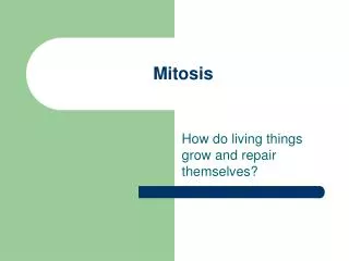
Overview Mitosis • Mitosis: • Purpose: • growth and repair in multicelled organisms • asexual reproduction in many organisms
Overview Mitosis • Mitosis: • An exact copy of the cell’s DNA is made, the copies separated, and each copy is put in a new cell. • Mitosis occurs in somatic cells
Mitosis • One division. • 1 cell 2 cells (called daughter cells) • Daughter cells are genetically identical • Chromosome number does not change.
Mitosis • Terms • Mitosis = division of the cell’s DNA and nucleus • Cytokinesis = division of the cytoplasm (cell)
Mitosis • Chromosome described/diagrammed • Sister chromatids have identical DNA • Centromere • Kinetechore on centromere provides binding site for microtubules
Chromosome Structure Histone core is made up of 8 proteins A nucleosome is 2 wraps of DNA around a histone core Histone core is shown in greater detail
Cell Cycle • Start with a newly formed cell • Cell cycle describes the “life cycle” of a cell (page 154): • Interphase • G1, S, G2 or • G0 (not shown in text) • Path followed by cells that will not divide again • Mitosis • Cytokinesis
Cell Cycle • Interphase G 1 - period of cell growth S - DNA synthesis • An exact copy is made of each chromosome • Copies are joined at the ________ G 2 – cell prepares to divide • e.g. centrioles duplicate in animal cells
Mitosis • Mitosis (division of nucleus) follows interphase • 4 phases • Prophase • Metaphase • Anaphase • Telophase
Prophase see pages 156/157 • Chromosomes condense, become visible under microscope • Centrioles move towards poles (animal only) • Nucleoli disappear • Nuclear envelope breaks up and forms vesicles
Plant Prophase • Early prophase in a plant cell • How would animal cell prophase differ from this?
Metaphase • Microtubules* attach sister chromatids to opposite poles (to centrioles in animal) • MT push and pull chromo to middle of cell • MT running pole to pole elongate cell • *MT arranged as spindle fibers Animal Metaphase
Plant Metaphase • Chromosomes tend to be “messier” in plant metaphase
Anaphase • Sister chromatids separate at centromere • MT pull sister chromatids to opposite poles • MT continue to elongate cell • This also helps to separate chromatids • Animal anaphase
Plant Anaphase • Separated sister chromatids clearly visible • No ________
Telophase and Cytokinesis • Telophase starts when chromatids reach poles • Goal is to make 2 new nuclei • Chromo. unwind • Nucleoli reappear • Nuclear envelope reforms from vesicles • _______ shown
Cytokinesis • Cytokinesis – division of cytoplasm • Begins during telophase • Different in plant and animal cells
Animal Cytokinesis • Microfilaments wrap around the center of the cell and then contract • Creates cleavage furrow • Cell “squeezed” in 2 Page 158
Plant Cytokinesis • Vesicles containing cell wall material line up across middle of cell • Vesicles merge and form cell plate • Cell plate grows until it divides the cell in 2 Cell plate
Mitosis Review • Comparison Plant and Animal Mitosis • Mitosis • Animal Cell Mitosis • Plant Cell Mitosis
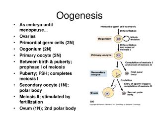
Meiosis • Goal of meiosis is to separate homologous chromosomes and produce gametes • Homologous Chromosomes: pair of chromosomes with genetic information about the same traits
Overview Meiosis • Meiosis: • Purpose of meiosis is to create gametes • Egg and sperm in humans • Needed for sexual reproduction • Occurs in germ cells • Ovaries and testes of humans
MEIOSIS • More on homologous chromosomes - see human karyotype on page 165
Meiosis • The process of meiosis requires 2 cellular divisions • Chromosomes are duplicated prior to only the 1st division
Related Terms • Diploid cell = 2 copies of each type of chromosome present (chromo. # = 2N) • One copy came from mom’s egg and the other from dad’s sperm • Human diploid number = 46 (also say 2N = 46) • Haploid cell = 1 copy of each type of chromosome present (chromo. # = N) • Human haploid number = 23 (N = 23)
Meiosis I • Chromosomes duplicate prior to meiosis I • Meiosis I – see page 166 • Prophase I • Metaphase I • Anaphase I • Telophase I
Meiosis I • Prophase I – remember chromo. are duplicated • Duplicated chromosomes form tetrads • Tetrad = pair of homologous chromosomes • Crossing over may occur • Homologous chromosomes exchange DNA segments – see board • Creates new combinations of DNA – creates chromosomes that are a combination of your mother’s DNA and your dad’s DNA
Meiosis I • Prophase I, continued • Chromosomes condense • Centrioles move towards opposite poles (animal only) • Spindle fibers begin to assemble • Nuclear envelope breaks down (always signals end of a prophase )
Meiosis I • Metaphase I • Spindle fibers attach the homologous chromosomes to opposite poles/centrioles • Spindle fibers push and pull the tetrads to the middle of the cell.
Meiosis I • Anaphase I • Homologous chromosomes are separated and pulled to opposite poles by the spindle fibers • Spindle fibers are made up of microtubules • Microtubules running pole to pole lengthen and elongate the cell
Meiosis I • Telophase I and Cytokinesis • Cell divides in two • Animal cells - cleavage furrow squeezes cell in two • Plant cells – cell plate divides cell in two • Nucleus does not reform
Meiosis I • At the end of meiosis I: • Homologous chromosomes have been separated • Chromosomes are still duplicated • Sister chromatids are no longer identical if crossing over has occurred • Chromosome number has been cut in half (to haploid number) • Count centromeres to count chromosomes
Meiosis II • Prophase II – in each cell • Centriole pairs separate and move to opposite poles (animal only) • Spindle fibers attach to kinetechore (centromere) of each chromosome • Remember chromosomes are still duplicated • Notice that each chromo is attached to both poles (as in mitosis)
Meiosis II • Metaphase II • Spindle fibers push and pull duplicated chromo. To the center of the cell
Meiosis II • Anaphase II • Spindle fibers pull the sister chromatids apart • One copy of each chromo moves to each pole • Microtubules running pole to pole lengthen and elongate the cell
Meiosis II • Telophase II and Cytokinesis • Nucleus reforms in each cell (4 cells in total) • Cytoplasm divides • Meiosis web link
Meiosis II • End result of meiosis • 4 cells made • Each cell has the haploid number of chromo. • One copy of each type of chromo • No two germ cells are identical due to: • independent assortment of homologous chromosomes • crossing over during meiosis I
Meiosis • Two cellular/nuclear divisions • 1st division separates homologous chromosomes (each in its duplicated state) • 2nd division separates duplicated chromosomes • 1 cell with 2N chromo 2 cells with N duplicated chromo 4 cells with N chromo
Meiosis Review • Meiosis
Cell Division Summary • Pages 172 and 173 provide a terrific summary of the 2 types of cellular division. • Given a picture of a phase of mitosis or meiosis you should be able to: • Identify the phase and division type • Label as appropriate: spindle fibers, centrioles, sister chromatids, homologous chromosomes, centromere/kinetechore, nuclear envelope…
Genetics Terms - Lab • Homozygous • Heterozygous • Genotype • Phenotype • Dominant • Recessive
Genetics • Recessive disorders (covered in lab) • Co-dominant alleles – blood type (covered in lab)
Genetics • Dominant disorders • Example: Huntington’s disease is dominant to being healthy
Genetics • Sex-linked disorders • X-linked: genetic information for the trait is located on the X chromosome • no genetic info about the trait on the Y chromo • Examples: Hemophilia and colorblindness • Y-linked (not many known) • Allele for hairy ears is y-linked