Download

1 / 42
520 likes | 1.38k Views
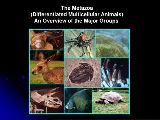
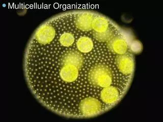
Origins of Multicellular Animals. Three Hypotheses. Syncytial ciliate hypothesis Ancestor is single celled ciliate with multiple nuclei Colonial flagellate hypothesis Ancestor is colonial flagellate like Volvox Polyphyletic hypothesis There may be multiple ancestors. THE PHYLUM PORIFERA.

E N D
Three Hypotheses • Syncytial ciliate hypothesis • Ancestor is single celled ciliate with multiple nuclei • Colonial flagellate hypothesis • Ancestor is colonial flagellate like Volvox • Polyphyletic hypothesis • There may be multiple ancestors
INTRODUCTION TO PORIFERA • unusual animals • originally thought they were plants • Are primarily marine, mostly in shallower waters • Are sessile and attached to substrate or objects- occasionally on other animals such as crabs • Are the most primitive metazoans and have neither true tissues or organs
Sponge Cell Types • Pinacocytes- outer cells; equivalent of epiderm
Sponge Anatomy Porocyte Amoebocyte Pinacocyte Choanocyte Pechenik, 1996
Sponge Cell Types • Pinacocytes- outer cells covering sponge; equivalent of epidermis • Choanocytes- • similar to choanoflagellates • collared cells with flagella - create water current and collect food matter.
Sponge Anatomy Porocyte Amoebocyte Pinacocyte Choanocyte Pechenik, 1996
Sponge Cell Types • Pinacocytes- outer cells covering sponge; equivalent of epiderm • Choanocytes- • similar to choanoflagellates • collared cells with flagella - create water current and collect food matter • Amoebocytes- • amoeba-like cells • store, digest and transport food, excrete wastes, secrete skeleton • give rise to buds in asexual reproduction
Mesophyl(=Mesenchyma) • Beneath the pinacocytes - a gelatinous protein layer • it contains the skeletal material (ie. spongin and spicules) and amoebocytes
Sponge Anatomy Porocyte Amoebocyte Pinacocyte Choanocyte Pechenik, 1996
Types of Spicules4 general types • Monaxon- needle-like or rod-like; straight or curved
Types of Spicules4 general types • Monaxon- needle-like or rod-like; straight or curved • Tetraxon- has 4 prongs
Types of Spicules4 general types • Monaxon- needle-like or rod-like; straight or curved • Tetraxon- has 4 prongs • Triaxon or Hexaxon- 3 or 6 rayed • Polyaxon- multiple short rods radiating from a common center; burr shaped, star shaped or like a child's jack. • Some species have a mixture of types
SponginGive phylum its common name • Some species have no spicules, but do have spongin • spongin is a type of hardened secreted protein • Some species have both spicules and spongin
Three Basic Sponge Types • Asconoid • Syconoid • Leuconoid
Asconoid Sponges • most primitive and simplistic in structure • have radial symmetry • are tube shaped
Asconoid Spongetwo basic openings • Ostia- • incurrent pores that open into a central cavity called the spongocoel • it is lined with choanocytes or collar cells • Osculum • the opening of the spongocoel to the outside • water leaves the sponge
Asconoid Sponge Design • Imposes definite size limits to sponges due to the problem of water flow • The spongocoel contains such a large volume of water that it is hard to push it out rapidly
Syconoid Sponges • next level of complexity • walls are invaginated • allowing for greater surface area over which water can pass • typically vase shaped like the asconoid sponges • radial symmetry
Syconoid Structure • helps to rectify some of the water movement problem • increasing the surface area • so there are more choanocytes to water volume • decreasing the spongocoel volume • these sponges able to get bigger than asconoid
Leuconoid Sponges • highest level of complexity in sponges • lost radial symmetry and are very irregular in shape and may attain large sizes • invaginated canals are even further invaginated and folded to from small flagellated chambers
Leuconoid Sponge Design • further increase in surface area makes these sponges highly efficient in moving and filtering water • spongocoel is gone except for canals that lead to the osculum- or there may be a series of excurrent openings • the largest sponges; most hydrologically efficient
Sponge Reproduction • Sexual • Asexual
Sexual Reproduction in Sponges • gametes formed by amoebocytes • there are both hermaphroditic and dioecious species • most hermaphroditic species produce eggs and sperm at different times so they do not self fertilize • sperm is released into environment via osculum and is brought in by another sponge via ostia • fertilization takes place in parent sponge • zygote is expelled - it drops to bottom and begins to develop
Asexual Reproduction in Sponges • two types: • Budding- fragmentation of body wall, buds appear as outgrowth on sides of sponge • when they reach a certain size they drop off and settle to bottom to form a new sponge • Gemmules- occurs only in freshwater sponges • gemmules are groups of food laden amoebocytes that deposit a hard covering of spicules around them • formation is triggered by environmental conditions such as decreased temperatures • they allow the sponge to pass the winter or periods of drought • after which the outer covering breaks open and a new sponge develops
Osmoregulation and Excretion in Sponges • no special organs • main waste is ammonia • it is removed by water currents within the sponge
HIGHER CLASSIFICATION OF SPONGES 4 classes of sponges
Class Calcarea • spicules composed of calcium carbonate • spicules are monaxons or tri or quadraxons • all three types of sponges exhibited • All less than 10 cm high • ex. Leucosolenia and Grantia • found in shallow coastal waters • all are marine
Class Hexactinellida(glass sponges) • Spicules of Silica and fused to form a lattice like skeleton • cup or vase shaped with well developed spongocoel • most beautiful example is Euplectella - venus flower basket • chiefly live in 500-1000 meter depth • are syconoid sponges • all are marine • may have commensal relationship with shrimp - where a male and female live inside the sponge; get trapped inside when they out-grow the pores of sponge
Class Demospongiae • Largest class - 95% of sponges in this class • spicules are silicious if present otherwise skeleton is made of spongin • variously shaped some are huge • all are leuconoid • all but one family is marine- Spongillidae- is freshwater about 150 freshwater species • this is the group from which we get our commercial sponges
ClassSclerospongiae • proposed in 1970 to include 6 species from Jamaica • have silicious spicules and spongin • also have an outer covering composed of calcium carbonate • are leuconoid sponges