Download
1 / 25
340 likes | 886 Views
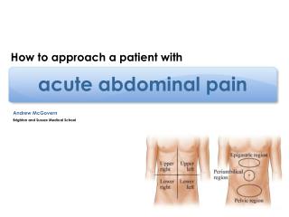
ACUTE ABDOMINAL PAIN. Abdul.Kader WEISS M.D CHIRURGIE GENERALE ET VISCERALE /CHIRURGIE COELIOSCOPIQUE D.E.S , A.F.S , A.F.S.A , DU / FRANCE Reference : Editors: Li Ern Chen , Timothy G. Buchman Title : Washington Manual of Surgery, The 5th Edition 2008 Lippincott Williams & Wilkins.

E N D
ACUTE ABDOMINAL PAIN Abdul.Kader WEISS M.D CHIRURGIE GENERALE ET VISCERALE /CHIRURGIE COELIOSCOPIQUE D.E.S , A.F.S , A.F.S.A , DU / FRANCE Reference : Editors: Li Ern Chen , Timothy G. Buchman Title: Washington Manual of Surgery, The 5th Edition 2008 Lippincott Williams & Wilkins
ACUTE ABDOMINAL PAIN • DEFINITIONS • Acute abdominal pain is the most common general surgical problem presenting to the emergency department. • ACUTE ABDOMEN IS DEFINED AS A RECENT OR SUDDEN ONSET OF ABDOMINAL PAIN. • This can be new pain or an increase in chronic pain. • The differential diagnosis includes both intra- and extraperitoneal processes. • The acute abdomen does not always signify the need for surgical intervention; however, surgical evaluation is warranted.
KEY POINTS • The level of abdominal pain generally relates to the origin: foregut- upper, midgut-middle, hindgut-lower. • Generally, colicky (visceral) pain is caused by stretching or contracting a hollowviscus(e.g. gallbladder, ureter, ileum). • Generally, constant localized (somatic) pain is caused by peritoneal irritation and indicates the presence of inflammation/infection (e.g. pancreatitis, cholecystitis, appendicitis). • Associated back pain suggests retroperitoneal pathology(aorticaneurysm, pancreatitis, posterior DU, pyelonephritis). • Associated sacral or perineal pain suggests pelvic pathology (ovarian cyst, PID, pelvic abscess). • Generally, very severe pain indicates ischaemia or generalized peritonitis(e.g. mesentericinfarction, perforatedduodenalulcer). • PAIN OUT OF PROPORTION TO THE PHYSICAL SIGNS SUGGESTS ISCHAEMIA WITHOUT PERFORATION. • REMEMBER REFERRED CAUSES OF PAIN: PNEUMONIA (RIGHT LOWER LOBE), MYOCARDIAL INFARCTION, LUMBAR NERVE ROOT PATHOLOGY.
PATHOPHYSIOLOGY • The abdomen is analogous to a box. • Although this chapter focuses on pathophysiology inside the box, one must be cognizant of the fact that pathology on the surface of the box (e.g., rectus sheath hematoma) or even outside the box (e.g., testicular torsion) can present as abdominal pain. • Abdominal pain arising from intra-abdominal pathophysiology originates in the peritoneum, which is a membrane comprising two layers. • These layers, the visceral and parietal peritoneum, are developmentally distinct areas with separate nerve supplies.
VISCERAL PAIN • Visceral peritoneum is innervated bilaterally by the autonomic nervous system. • The bilateral innervation causes visceral pain to be midline, vague, deep, dull, and poorly localized (e.g., vague periumbilical pain of the midgut). • Visceral pain is triggered by inflammation, ischemia, and geometric changes such as distention, traction, and pressure. • Visceral pain signifies intra-abdominal disease but not necessarily the need for surgical intervention.
PARIETAL PAIN • Parietal peritoneum is innervated unilaterally via the spinal somatic nerves that also supply the abdominal wall. • Unilateral innervation causes parietal pain to localize to one or more abdominal quadrants (e.g., inflamed appendix producing parietal peritoneal irritation). • Parietal pain is sharp, severe, and well localized. • Parietal pain is triggered by irritation of the parietal peritoneum by an inflammatory process (e.g., chemical peritonitis from perforated peptic ulcer or bacterial peritonitis from acute appendicitis). • It may also be triggered by mechanical stimulation, such as a surgical incision. • Parietal pain is associated with physical examination findings of local or diffuse peritonitis and frequently signifies the need for surgical treatment.
EMBRYOLOGIC ORIGIN • Embryologic origin of the affected organ determines the location of visceral pain in the abdominal midline. • Foregut-derived structures (stomach to the second portion of the duodenum, liver and biliary tract, pancreas, spleen) present with epigastric pain. • Midgut-derived structures (second portion of the duodenum to the proximal two thirds of the transverse colon) present with periumbilical pain. • Hindgut-derived structures (distal transverse colon to the anal verge) present with suprapubic pain.
REFERRED PAIN • Referred pain arises from a deep visceral structure but is superficial at the presenting site . • It results from central neural pathways that are common to the somatic nerves and visceral organs. • Examples include biliary tract pain (referred to the right inferior scapular area) and diaphragmatic irritation from any source, such as subphrenic abscess (referred to the ipsilateral shoulder).
REFERRED PAIN Solid circles are primary or most intense sites of pain Solid circles are primary or most intense sites of pain
EVALUATION • Evaluation of the acute abdomen remains heavily influenced by patient history and physical exam findings. • Ancillary imaging and lab tests can help to complete the diagnosis and guide treatment decisions.
EVALUATION • HISTORY OF PRESENT ILLNESS • PAST MEDICAL HISTORY, SURGICAL HISTORY, AND ORGAN-SYSTEM REVIEW. • MEDICATIONS • PHYSICAL EXAMINATION • LABORATORY EVALUATION • RADIOLOGIC EVALUATION
HISTORY OF PRESENT ILLNESS • ONSET AND DURATION OF PAIN • Sudden onset of pain (within seconds) suggests perforation or rupture [e.g., perforated peptic ulcer ]. • Rapidly accelerating pain (within minutes) may result from several sources , such as colic syndromes, [ e.g. biliary colic ] , Inflammatory processes [ e.g. acute appendicitis ] ,Ischemic processes [ e.g. mesenteric ischemia ] . • Gradual onset of pain (over several hours) increasing in intensity may be caused by one of the following: • Inflammatory conditions, such as appendicitis and cholecystitis. • Obstructive processes, such as nonstrangulated bowel obstruction and urinary retention. • CHARACTER OF PAIN • Colicky pain waxes and wanes. It usually occurs secondary to hyperperistalsis of smooth muscle against a mechanical site of obstruction (e.g., small-bowel obstruction, renal stone). • An important exception is biliary colic, in which pain tends to be constant. • Pain that is sharp, severe, persistent, and steadily increases in intensity over time suggests an infectious or inflammatory process (e.g., appendicitis). • LOCATION OF PAIN • Pain caused by inflammation of specific organs may be localized [e.g., right-upper-quadrant (RUQ) pain caused by acute cholecystitis]. • Careful attention must be given to the radiation of pain. The pain of renal colic, for example, may begin in the patient's back or flank and radiate to the ipsilateral groin, whereas the pain of a ruptured aortic aneurysm or pancreatitis may radiate to the patient's back. • ALLEVIATING AND AGGRAVATING FACTORS • ASSOCIATED SYMPTOMS • Nausea and vomiting frequently accompany abdominal pain and may hint at its etiology. Vomiting that occurs after the onset of pain may suggest appendicitis, whereas vomiting before the onset of pain is more consistent with the diagnosis of gastroenteritis or food poisoning. • Fever or chills suggests an inflammatory or an infectious process, or both. • Anorexia is present in the vast majority of patients with acute peritonitis.
CHARACTER OF PAIN GRADUAL, PROGRESSIVE PAIN COLICKY, CRAMPY, INTERMITTENT PAIN
SUDDEN, SEVERE PAIN CHARACTER OF PAIN
B. PAST MEDICAL HISTORY, SURGICAL HISTORY, AND ORGAN-SYSTEM REVIEW • Pathologic medical conditions may precipitate intra-abdominal pathology. • Patients with peripheral vascular disease or coronary artery disease may have abdominal vascular disease (e.g., AAA or mesenteric ischemia). • Patients with a history of cancer may present with bowel obstruction from recurrence. • Major medical problems are important to recognize early in the patient and may call for urgent surgical exploration. • A thorough medical history and organ-system review must be carried out to exclude various extra-abdominal causes of abdominal pain. • Diabetic patients or patients with known coronary artery disease or peripheral vascular disease who present with vague epigastric symptoms may have myocardial ischemia as the cause of the abdominal symptoms. • Right-lower-lobe pneumonia may present as RUQ pain in association with cough and fever. • A thorough menstrual history must be obtained in women. • Pelvic inflammatory disease (PID) typically occurs early in the cycle and may be associated with a vaginal discharge. • Ectopic pregnancy must be considered in every woman of child-bearing age with lower abdominal pain, especially if accompanied by a history of amenorrhea. • Abdominal pain that occurs monthly suggests endometriosis. • Previous abdominal surgery in a patient with colicky abdominal pain may suggest intestinal obstruction secondary to adhesions, incarceration of an incisional hernia, or recurrence or malignancy. These are generally accompanied by nausea and vomiting.
C. MEDICATIONS • NONSTEROIDAL ANTI-INFLAMMATORY MEDICATIONS, such as aspirin or ibuprofen, place patients at risk for the complications of peptic ulcer disease, including bleeding, obstruction, and perforation. • CORTICOSTEROIDS may mask classic signs of inflammation, such as fever and peritoneal irritation, making the abdominal examination less reliable. • ANTIBIOTICS consumed by patients may aid or hinder diagnosis. • Patients with peritonitis may have decreased pain. • Patients who have diarrhea and abdominal pain may have antibiotic-induced pseudomembranous colitis caused by Clostridium difficile. • Be aware of the elderly patient on immunosuppressants or antibiotics.
D. PHYSICAL EXAMINATION • OVERALL APPEARANCE SHOULD BE ASSESSED (e.g. jaundice ) • VITAL SIGNS are important indicators of a patient's overall condition ( Bp , P , T ) . • THE ABDOMINAL EXAMINATION should be carried out thoroughly and systematically. • INSPECTION ( distention, surgical scars, bulging masses, and areas of erythema ). • AUSCULTATION ( high-pitched, tinkling bowel sounds of obstruction , absence of sounds due to ileus from diffuse peritonitis ). • PERCUSSION ( tympanitic sounds of distended bowel in intestinal obstruction , fluid wave that is characteristic of ascites ). • PALPATION of the patient's abdomen should be performed with the patient in a supine position and with his or her knees flexed, if necessary, to relieve pain. • RECTAL EXAMINATION should be performed routinely in all patients with abdominal pain. • Tenderness or a mass on the right pelvic side wall is sometimes seen in appendicitis. • A mass in the rectum may indicate obstructing cancer. • The presence of occult blood in the stool specimen may indicate GI bleeding from peptic ulcer disease. • PELVIC EXAMINATION must be performed in all women of child-bearing age who present with lower abdominal pain. • TESTICULAR AND SCROTAL EXAMINATION is essential in all males who complain of abdominal pain ( Testicular torsion , Epididymitis ) . • SPECIFIC PHYSICAL EXAMINATION findings should be sought in the appropriate clinical setting. • Murphy's sign is inspiratory arrest while continuous pressure is maintained in the RUQ , seen in acute cholecystitis . • Rovsing's seen in acute appendicitis. Indicative of an inflammatory process in the right lower quadrant (RLQ), Rovsing's sign is RLQ pain resulting from percussion in the left lower quadrant (LLQ).
E. LABORATORY EVALUATION • A complete blood count with cell count differential • White blood cell (WBC) count elevation may indicate the presence of an infectious source. • Hematocrit elevation may be due to volume contraction from dehydration. Conversely, a low hematocrit may be due to occult blood loss. • An electrolyte profile may reveal clues to the patient's overall condition. • Hypokalemic, hypochloremic, metabolic alkalosis may be seen in patients with prolonged vomiting and severe volume depletion. • Elevation of the blood urea nitrogen or creatinine is also indicative of volume depletion. • Liver enzyme levels may be obtained in the appropriate clinical setting. • A mild elevation of transaminases (<2 times normal), alkaline phosphatase, and total bilirubin is sometimes seen in patients with acute cholecystitis. • A moderate elevation of transaminases (>3 times normal) in the patient with acute onset of RUQ pain is most likely due to a common bile duct (CBD) stone. • Pancreatic enzymes (amylase and lipase) should be measured if the diagnosis of pancreatitis is considered. • Mild degrees of hyperamylasemia may be seen in several situations, such as intestinal obstruction. • Elevation of lipase usually indicates pancreatic parenchymal damage. • Lactic acid level may be obtained when considering intestinal ischemia. • Serum lactate is an indicator of tissue hypoxia. • Mild lactic acidosis may be seen in patients with arterial hypotension. • Urinalysis is helpful in assessing urologic causes of abdominal pain. • Bacteriuria, pyuria, and a positive leukocyte esterase usually suggest a urinary tract infection (UTI). • Hematuria is seen in nephrolithiasis and renal and urothelial cancer. • β-Human chorionic gonadotropin must be obtained in any woman of child-bearing age. A positive urine result should be quantitated by serum levels.
F. RADIOLOGIC EVALUATION : its use should be very selective to avoid unnecessary cost and possible morbidity associated with some modalities. • Plain abdominal x-rays often serve as the initial radiologic evaluation. X-rays should be obtained in the supine and erect positions. • Ultrasonography (US) may provide diagnostic information in some conditions. Ultrasound is portable, relatively inexpensive, and free of radiation exposure. US visibility is limited in settings of obesity, bowel gas, and subcutaneous air. • RUQ US is particularly useful in biliary tract disease. • US can be used in the evaluation of RLQ pain. • Contrast studies, although rarely indicated in the acute setting, may be helpful in some situations. • In most instances, a water-soluble contrast agent (e.g., Hypaque) should be used to avoid possible barium peritonitis in the event of bowel perforation. • Contrast enema is particularly useful in differentiating adynamicileus from distal colonic obstruction. • Computed tomographic (CT) scanning may provide a thorough evaluation of the patient's abdomen and pelvis relatively quickly. Oral and intravenous contrast should be administered if not specifically contraindicated by allergy, renal insufficiency, or patient hemodynamic instability. CT scanning is the best radiographic study in the patient with unexplained abdominal pain. • Magnetic resonance imaging (MRI) • MRI provides cross-sectional imaging while avoiding ionizing radiation. • Image acquisition takes longer than for CT scan; patients must be able to lie on their backs for a prolonged period of time and cannot be claustrophobic. • MRI has its greatest application in pregnant women with acute abdominal and pelvic pain . • Radionuclide imaging studies have few indications in the acute setting.