Download
1 / 44
440 likes | 584 Views
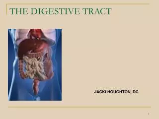
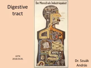
I. BASIC FUNCTIONS OF THE DIGESTIVE TRACT. A. Digestion —process of altering the physical state and chemical composition of food so that the body’s cells can use it

E N D
I. BASIC FUNCTIONS OF THE DIGESTIVE TRACT A. Digestion—process of altering the physical state and chemical composition of food so that the body’s cells can use it B. Absorption—process by which small digested molecules pass through the cells of the intestinal tract, entering the blood and lymph
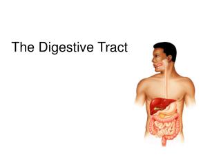
II. ANATOMY OF DIGESTIVE SYSTEM A. Components 1. Alimentary Canal • Mouth • Pharynx • Esophagus • Stomach • Small intestine • Large intestine 2. Accessory Organs • Salivary glands • Liver • Gallbladder • Pancreas
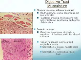
B. Wall Structure of Alimentary Canal 1. Alimentary canal is a muscular tube, 30 feet long, and located in the ventral body cavity. • Has the same four layers throughout: a. Mucosa • Innermost layer b. Submucosa • Loose CT with blood vessels, glands, lymph vessels, and nerves c. Muscularis mucosa • 2 layers of smooth muscle d. Serosa • Outermost layer (visceral peritoneum)
C. Movement of the tube • 2 Basic Movements: 1. Mixing—mixes food with juices secreted by the mucosa of the stomach 2. Propelling movements—peristalsis (wavelike contractions that force food along the digestive tube) Mixing Peristalsis
D. Oral Cavity 1. Mouth • Receives food • Prepares food for digestion (breaks food into small particles and mixes it with saliva) 2. Tongue • Mostly muscle • Anchored to midline of the floor of the mouth by the frenulum • Covered with papillae which contain taste buds • Movement aids in mixing food and saliva and moving food toward the rear of the mouth
3. Palate • Forms roof of the mouth • Consist of hard anterior part (hard palate) and soft posterior part (soft palate) • Uvula—cone-shaped projection that hangs down from soft palate and pulls upward when swallowing to prevent food from entering the nasal cavity 4. Tonsils • Masses of lymphatic tissue • 3 tonsil masses: a. palatine b. pharyngeal (adenoids) c. lingual
5. Teeth • 2 sets a. primary(deciduous)—20 b. secondary (permanent)—32 • F(x): mastication (chewing) • 4 types of teeth: a. incisors—front teeth for biting b. canine—cone-shaped for tearing food c. bicuspids—for grinding food particles d. molars—for grinding food particles
Consists of: • crown—part above gum • root—anchored to bone by cementum and periodontal ligament • enamel—covers crown --hardest substance in body d. dentin—under enamel --like very hard bone e. pulpcavity—under dentin --contains blood vessels, nerves, and CT
E. Salivary Glands 1. F(x): • To secrete saliva which moisten food and begins carbohydrate digestion • Cleanses mouth and teeth • Dissolves food for taste 2. Consists of serous cells which produce amylase (enzyme that breaks down starch and glycogen) and mucous cells which secrete mucus for lubrication 3. 3 major pairs of salivary glands a. parotids—in front of and below each ear b. submandibular—in floor of mouth c. sublingual—on floor of mouth under tongue
F. Pharynx 1. Common to digestive and respiratory tracts 2. Divided into 3 areas: a. nasopharynx—passage for air during breathing b. oropharynx—passageway for food and air c. laryngopharynx—opens into larynx and esophagus 3. F(x): • Swallowing (deglutition) -Voluntary but becomes involuntary as swallowing reflex is initiated -Involves chewing and bolus (ball of partially digested food) formation
G. Esophagus 1. Collapsed tube about 10 inches long that connects pharynx and stomach 2. Mucous glands keep it moist and lubricated H. Stomach 1. Anatomy • “J” shaped pouch-like organ just under the diaphragm in the upper left portion of abdominal cavity • Inner mucosa forms folds called rugae
2. F(x): • Receive food • Mix food with gastric juice • Initiate protein digestion • Limited absorption • Transport partially digested food to small intestine 3. Divided into 4 regions: a. cardiac—near esophageal opening b. fundus—temporarystorage area c. body d. pylorus—enters small intestine
4. Mucosa is thick with many gastric glands. 5. Gastric glands contain 3 types of secretory cells: a. goblet cells—secrete mucus b. chief cells—secrete digestive enzyme- pepsinogen (inactive form of pepsin which digest proteins) c. parietalcells—secrete hydrochloric acid (HCl) and intrinsic factor
6. F(x) of gastric gland secretions: a. mucus—protection b. HCl—converts pepsinogen to pepsin c. intrinsicfactor—aids in absorption of vitamin B12 in small intestine 7. Regulation of Gastric Secretion • Under nerve and hormone control • Gastrin(stomach hormone) increases release of gastric juices 8. Substances absorbed in stomach: • water • glucose • alcohol • aspirin • lipid-soluble drugs
9. Mixing and Emptying of Stomach • Mixing produces chyme(semisolid paste) and peristalsis moves it to the pylorus • Rate of emptying depends on the type of food present • Liquids pass through rapidly • Solids remain until well mixed with gastric juices • Fatty food remains the longest • Carbohydrates pass through the fastest
PANCREAS A. Structure • Elongated, flattened organ • Extends horizontally across the posterior abdominal wall in the C-shaped curve of the duodenum • Pancreatic secretions enter the duodenum(small intestine) through the pancreatic duct 4. Heterocrine Gland (endocrine and exocrine) 5. Exocrine Component functions in digestion • Pancreatic acinar cells -make up most of the pancreas -produce pancreatic juice
LIVER A. Structure 1. Macroscopic • Reddish-brown in color • Enclosed in a fibrous capsule • Well supplied with blood vessels • Located in the upper right side of the abdominal cavity inferior to the diaphragm • Divided into 2 lobes (large right lobe and smaller left lobe) 2. Microscopic • Each lobe is separated into many tiny hepatic lobules (functional unit of liver) • Each lobule has many hepatic (cuboidal) cells radiating outward from a central vein
Functions of the Liver • Carries out the metabolism of carbohydrates, lipids, and proteins • Storage • Stores glycogen (animal starch), iron, blood, and vitamins A, D, B12 3. Blood filtering • Removal of damaged red blood cells and foreign substances
III. GALLBLADDER A. Structure 1. Pear-shaped sac on the inferior surface of the liver 2. Lined with epithelial cells 3. Wall contains a strong, muscular layer 4. Connects to cystic duct which joins with the common hepatic duct to form the common bile ductwhich empties into the duodenum B. Functions • Store bile • Concentrate bile by reabsorbing water • Release bile into small intestine
Gallstones 1. Crystals formed from cholesterol in bile 2. Can block bile flow, cause pain, and result removal of gallbladder
SMALL INTESTINE A. Structure • Tubular organ about 20 feet long • Joins the stomach at the pyloric sphincter • Joins the large intestine at the ileocecal junction • 3 Divisions: • duodenum~10 inches long and 2 in. in diameter (fixed) • jejunum~8 feet long • ileum~12 feet long 5. Jejunum and ileum are suspended by the mesentery (tissue containing blood vessels, nerves, and lymphatic vessels that supply the intestinal wall)
Functions of the Small Intestine 1. Completes digestion 2. Absorbs products of digestion 3. Receives secretions from pancreas and liver 4. Transports residues to large intestine
V. LARGE INTESTINE A. Structure • 5 feet long • 4 Divisions of Large Intestine a. Cecum • First 2-3 inches • Blind pouch to which is attached the vermiform appendix (lymphatic tissue which has no digestive function) • Opening between ileum and cecum controlled by ileocecal valve
b. Colon (4 parts) • Ascending (right side) • Transverse (longest part) • Descending (left side) • Sigmoid (S-shaped)
c. Rectum • From colon to anal canal d. Anal Canal • Last 1-2 inches of large intestine • Anus -opening on distal end of anal canal -controlled by 2 sphincter muscles: internal anal sphincter-- involuntary external anal sphincter-- voluntary
B. Functions of the Large Intestine • secrete mucus • reabsorb water and electrolytes • store and eliminate waste
C. Feces • Solid waste • Undigested or unabsorbed material • 75% water • Color due to bile pigments • Odor from bacterial activity