Download
1 / 27
270 likes | 468 Views
BAV completo: Dissociazione AV (pacemakers autonomi nodo seno e ventricoli). Dissociazione AV. Dissociazione AV. QRS normale. Durata: 0.06"-0.10" Q non superiore a 0.04" in D'1, D2, aVL, aVF Q puo' essere > 0.04" in D3, aVR, V1, V2 Q in D1 e D2: non > 2 mm, non > 25% di R.

E N D
BAV completo: Dissociazione AV (pacemakers autonomi nodo seno e ventricoli)
QRS normale Durata: 0.06"-0.10" Q non superiore a 0.04" in D'1, D2, aVL, aVF Q puo' essere > 0.04" in D3, aVR, V1, V2 Q in D1 e D2: non > 2 mm, non > 25% di R In precordiali: voltaggio R sale da V1 a V5 voltaggio R in V6 > a Q o S V3, V4: zona transizione (complessi isodifasici) tra ventr dx (V1, V2) e ventr sin (V5, V6) V1-V2: intervallo tra inizio QRS ed apice R (R')< 0.03" V5-V6: intervallo tra inizio QRS ed apice R (R')< 0.05"
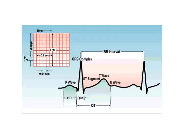
Question 2 of 10 What is the QRS duration seen here? A. 0.04 sec B. 0.06 sec C. 0.10 sec D. 0.12 sec E. 0.14
What potentially fatal abnormality can be seen in this ECG? A. Increased PR interval B. Poor R wave progression (V1 - V3) C. Increased QT interval D. Decreased QT interval E. Increased P wave amplitude
Determine the QRS axis for this ECG:A. IndeterminateB. +90 degreesC. -30 degreesD. -45 degreesE. +150 degrees D1 neg aVF pos D2 isodif
Determine the QRS axis for this ECG:A. IndeterminateB. +90 degreesC. -30 degreesD. -45 degreesE. +150 degrees
Determine the axis for this ECG:A. +90 degreesB. IndeterminateC. +30 degreesD. -30 degreesE. -150 degrees D1 pos aVF pos D3 isodif
Determine the QRS axis for this ECG:A. -60 degreesB. -45 degreesC. +60 degrees D. IndeterminateE. 0 degrees D1 pos aVF isodif
aVF neg Determine the QRS axis for this ECG:A. -75 degreesB. -30 degreesC. 0 degreesD. +45 degreesE. Indeterminate D3 neg D2 neg aVR pos D1 pos
Determine the QRS axis for this ECG:A. -15 degreesB. +15 degreesC. +60 degreesD. +105 degreesE. Indeterminate axis Occasionally each of the 6 frontal plane leads is small and/or isoelectric. The axis cannot be determined and is called indeterminate. This is a normal variant
Determine the QRS axis for this ECG:A. -100 degreesB. -30 degreesC. +15 degreesD. +90 degreesE. Indeterminate aVF pos D1 isodif