Download
1 / 31
310 likes | 396 Views
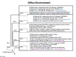
3D Tumor Stem Cell Niche Image Analysis. NCI-ICBP CMCD U54 Progress Report. Fuhai Li, Ph.D. BBP-TMHRI, Feb 7 2011. http://www.methodisthealth.com/icbp. Tumor Stem Cell Niche. J Rosen, BCM. From 2D to 3D. DAPI. GFP. From center grant proposal. Dextran. Superimposed.

E N D
3D Tumor Stem Cell Niche Image Analysis NCI-ICBP CMCD U54 Progress Report Fuhai Li, Ph.D. BBP-TMHRI, Feb 7 2011 http://www.methodisthealth.com/icbp
Tumor Stem Cell Niche J Rosen, BCM
From 2D to 3D DAPI GFP From center grant proposal Dextran Superimposed .44 μm X .44 μm X 1 μm
Specific Aim • Digitize 3D images of tumor stem cell niche & provide quantitative data for • Tumor modeling • Blood vessel tracing • Nuclei segmentation
What information is expected? • Anything we can see, image, and imagine! E.g., • Spatial distributions (in normal and tumor, before and after treatment) of blood vessels, stem cells, stroma, etc. • Cell morphology, proliferation and death From John, Tegy, Mei Zhang, Michael T. Lewis
Blood Vessel Segmentation (Tracing) Technical Challenges: Noise Signal Broken Points and Vessel Crossing
Validation of Available Segmentation Software http://research.mssm.edu/cnic/tools-ns.html
NeuronStudio http://research.mssm.edu/cnic/tools-ns.html
V3D http://penglab.janelia.org/proj/v3d/V3D/About_V3D.html
FARSIGHT http://www.farsight-toolkit.org/wiki/Main_Page
Multiscale Vessel Enhancement Filtering Multiscale means image filtering with Gaussian filters (with different sigmas) The eigenvalues of Hessian matrix tell us where are the vessels Hessian Matrix A. Frangi, et. al, MICCAI, 1998
Skeleton Extraction T.C. Lee, et. al, GVGIP, 1994
Binary 3D View
Optimal Linking by Integer Programming (ongoing) smoothness term Fitness term Cost function: Solving: subject to: can only be linked to at most to once!
Nuclei Segmentation Technical Challenges: Uneven Intensity and Cell Clustering
FARSIGHT 3D Analysis
Flowchart of Nuclei Segmentation Z-Series 2D Images Adaptive Thresholding Distance Transform (Shape Info.) LoG Filtering (Intensity Info.) Seed Image (Non-Maximum Suppression) Level Set Segmentation
TT Proposed 3D Analysis
Refine & Define Nuclei by Ellipsoid Fitting digital 3D tumor stem cell niche nucleus = {o, a, b, c, α, β} Low Z resolution
Ongoing Work • Blood vessel smoothing & broken points linking • Nuclei ellipsoid fitting, Image quantification & dividing cell identification • Process large volumes of image data to derive information for tumor modeling • Graphic user interface (GUI) tools for image analysis, visualization & tumor simulation
TT Normal Breast Tissue From Dr. Michael T. Lewis (John David Acquired)
Acknowledgements BCM: Michael Lewis Mei Zhang Mary Dickinson John Landua Tegy Vadakkan Wei Wei BBP-TMHRI: Stephen Wong Xiaofeng Xia Hong Zhao Derek Cridebring NIH NCI-ICBP U54 Grant Support