Download
1 / 24
240 likes | 410 Views
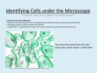
Microscope and Cells. Chapter 4. I. The Cell Theory. All living organisms are made up of cells Cells are the basic units of structure and function in living organisms. All cells come from cells that existed before them by cellular reproduction.

E N D
Microscope and Cells Chapter 4
I. The Cell Theory • All living organisms are made up of cells • Cells are the basic units of structure and function in living organisms. • All cells come from cells that existed before them by cellular reproduction. • Schwann, Schleiden and Virchow are credited with coming up with the basics of the cell theory • http://www.youtube.com/watch?v=dscY_2QQbKU
Every cell has the following main characteristics: • Plasma membrane covering • Cytoplasm • Their genes are made of DNA • Ribosomes are tiny organelles that assemble proteins
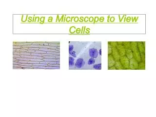
II. The Compound Light Microscope • Antone von Leeuwenhoek assembled the first microscope that was useful for scientific research. • Compound light microscopes reflect light through a set of lenses and the specimen to magnify the specimen. • See handout for the parts of the microscope – you must know it.
Two important characteristics that determine the quality of a light microscope: • Magnification – an increase in the apparent size of an object. We calculate magnification by the following: Magnification of eyepiece x magnification of objective = total magnifying power • Resolution – the measure of clarity of an image. As the magnification increases, the resolution of the image decreases.
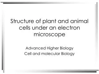
III. Electron Microscopes • Some microscopes use beams of electrons for magnification instead of light – electron microscopes • Scanning electron microscope (SEM) – used to study the detailed architecture of the surface of the object. Forms a 3D image, but does not show the inside of the object. • Transmission electron microscope (TEM) – used to provide a detailed 2D image of the inside structure of the object that is viewed.
http://micro.magnet.fsu.edu/primer/java/electronmicroscopy/magnify1/index.htmlhttp://micro.magnet.fsu.edu/primer/java/electronmicroscopy/magnify1/index.html • http://www5.pbrc.hawaii.edu/microangela/index.html
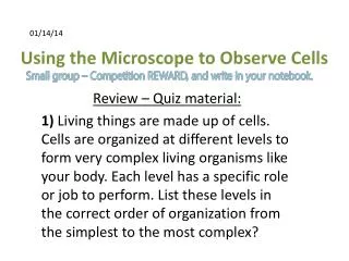
III. The Size of Cells • http://www.cellsalive.com/howbig.htm • Cells are microscopic, they are visible only with light microscopes. Most of their size ranges from 1-100 µm. • The cells are small, because they have to be able to carry materials from one side of the cell to the next in a short period of time. • Cells must have a large enough surface area to be able to take in nutrients and oxygen and release waste quickly.
IV. Prokaryotic Cells • Prokaryotic cells – small cells (about 1-10 µm) that do not have a nucleus and organelles that are covered with membranes • Prokaryotic cells are found in Bacteria and Archaea (two main domains of life).
Nucleoid region – part of the prokaryotic cell where the DNA is found • Cell membrane – innermost covering of the cell • Cell wall – made up of a special mix of polysaccharides and proteins (antibiotics break it down) • Capsule – outside of the cell wall, protective covering (not all bacteria have it) • Flagella (sing. Flagellum) – moves bacteria • Pili – used to stick them to surfaces and for conjugation (exchange of genetic materials between bacteria)
Cytoplasm – dissolves substances and holds organelles • Ribosomes – organelles that make proteins in the cytoplasm • http://www.ted.com/talks/lang/eng/bonnie_bassler_on_how_bacteria_communicate.html • http://www.youtube.com/watch?v=RGfs328X8Kw – artificial bacteria • http://www.youtube.com/watch?v=tqOVYpkZ0qs – bacteria song
V. Eukaryotic Cells • Protists, Fungi, Plants, and Animals • Have nucleus and organelles that are surrounded by phospholipid membranes (membrane-bound organelles) • Much larger and more complex than prokaryotic cells. (10-100 µm) • Reproduce sexually and asexually • http://www.youtube.com/watch?v=Y3u_GDIj7RI&feature=related
VI. Eukaryotic OrganellesGeneral Function: Manufacturing • Nucleus • Control center of cell; contains most of the cell’s DNA • Nucleolus • Location where ribosomes are synthesized, made up of DNA, RNA and proteins • Nuclear envelope • Protects the DNA in the nucleus, nuclear pores allow the exchange of materials. • Amazing cells: http://learn.genetics.utah.edu/content/begin/cells/insideacell/
Ribosomes • Protein synthesis – mushroom-shaped organelle that is made up of RNA and proteins • Rough ER • Comprised of a network of tubes and flattened sacs. • Continuous with plasma membrane and nuclear membrane and made up of the same material • Site of protein synthesis (consists of ribosomes) and protein folding (this is where many proteins get their secondary and tertiary structures).
Smooth ER • Site of lipid and carbohydrate metabolism • No ribosomes • Membrane structure, so it is also made up of phospholipids and proteins • Detoxification • Golgi Apparatus • Connected with ER; flattened disc-shaped sacs, stacked one on top of the other • Modification, storage, and packaging of proteins. • “Tags” proteins so they go to the correct destination • “The mailroom of the cell”
VII. Eukaryotic OrganellesGeneral Function: Breakdown • Lysosomes (in animal cells and some protists) • Digestion of nutrients, bacteria, and damaged organelles; destruction of certain cells during embryonic development • Peroxisomes • Diverse metabolic processes break down H ions into hydrogen peroxide • Detoxification • Vacuoles • Digestion (like lysosomes); storage of chemicals, cell enlargement; water balance, really large in older plant cells
Vesicles – small membrane bubbles that ship materials within and out of the cell.
VIII. Eukaryotic Organelles: Energy Processing • Chloroplasts • Conversion of light energy to chemical energy of sugars (site of photosynthesis) • Double membrane structure You must be able to draw and label the parts of chloroplasts • Mitochondria • Conversion of chemical energy of food to chemical energy – cellular respiration • Bound by double membrane You must be able to draw and label the parts of mitochondria http://multimedia.mcb.harvard.edu/anim_mitochondria.html
IX. Eukaryotic OrganellesGeneral Function: Support, Movement, and Communication • Cytoskeleton (including cilia, flagella, and centrioles in animal cells) • Maintenance of cell shape; anchorage for organelles; movement of organelles within cells; cell movement; mechanical transmission of signals from exterior of cell to interior. Cilia and flagella move the cell or move substances on the cell. • Cell walls (in plants, fungi, and protists) • Maintenance of cell shape and skeletal support; surface protection; binding of cells in tissues • Made up of cellulose in plants, chitin in fungi and different types of materials in prokaryotes.