Download
1 / 64
640 likes | 702 Views
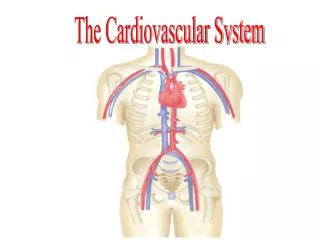
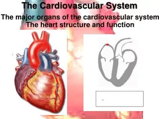
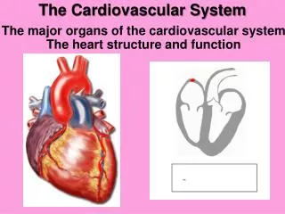
Cardiovascular System:. The Heart. Heart Size, Shape, Mass. About the size of a closed fist: 3.5” wide (at widest pt) X 5” long. 2.5” thick Cone-shaped: Base & Apex 8 oz in adult females, 10 oz in adult males. Heart Location. In the mediastinum (tissue between sternum & vertebral column)

E N D
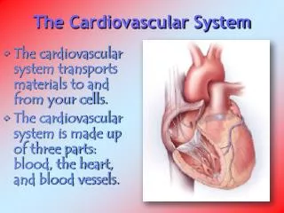
Cardiovascular System: The Heart
Heart Size, Shape, Mass • About the size of a closed fist: • 3.5” wide (at widest pt) X 5” long. 2.5” thick • Cone-shaped: Base & Apex • 8 oz in adult females, 10 oz in adult males
Heart Location • In the mediastinum (tissue between sternum & vertebral column) • 2/3 of its mass is left of midline • A cone lying on its side: • Base is toward your right shoulder, apex points to your left hip • Anterior surface - deep to sternum • Inferior surface – on diaphragm • Right border – against right lung. • Left border (pulmonary border) – against left lung
Fibrous, Serous (Visceral & Parietal) pericardium
Fibrous & Serous pericardium • Pericardium – the sac that surrounds, protects, anchors heart to diaphragm. It is composed of 2 layers - the fibrous & serous (visceral & parietal layers) pericardium
The FIBROUS Pericardium • FIBROUS PERICARDIUM • Outermost layer • Tough, inelastic, dense irregular CT • Prevents overstretching of heart • Anchors heart to diaphragm. Prevents rising of heart
The SEROUS PERICARDIUM SEROUS pericardium: thinner, delicate, inner layers that form a fluid-filled sac. It has 2 continuous layers: • Parietallayer: is adhered to fibrous pericardium • Visceral layer aka “Epicardium”: is adhered to heart • The pericardial cavity is filled with Pericardial fluid. The pericardial cavity is the space between parietal & visceral layers. • The pericardial fluid PREVENTS FRICTION
Systemic & Pulmonary Circulations • Two closed circulatory systems: • Body • Lungs • The output of one becomes the input of the other with each beat of the heart
Cardiac Output (CO) • Total volume of blood in the body is approximately 5L. CO is typically about 5L/min • CO= Amount of blood pumped by left or right ventriclePER MINUTE • CO depends on Heart Rate & Stroke Volume • HEART RATE ie number of beats per minute. Normal is 75BPM • STROKE VOLUME ie. the amount of blood ejected from one Ventricle PER BEAT. Normal is 70ml/beat in a 70 kg healthy man Heart pumps 5 L blood PER MINUTE
Autorhythmicvs Contractile myocytes • The Heart has 2 kinds of cells: • Autorhythmicmyocytes (purple circle / yellow cell) spontaneously depolarize and generate action potentials • Contractile myocytes(pink cells) contract together to pump blood as the action potential spreads across them
The Sinoatrial(SA) Node or Pacemaker • The SinoAtrial (SA) node is an area of modified cardiac myocytes within the R atrium. • Cells here spontaneously depolarize (become more positive) 100 x/min • Pacemaker potential: the spontaneous depolarization from -60 to -35mV that precedes the action potential • Action potential: the depolarization that occurs after threshold of -35 mV
Action Potential at the SA Node SA node cells do not “rest” • Na+channels spontaneously open at -60mV. (If –funny current) • Na+ leaks into SA node cellswhich initiates the pacemaker potential • 2 Ca2+ channels open. One at low voltage, one at high voltage. • Ca2+ enters the cell, causing part of pacemaker potential & then the action potential • At 0mV, K+ channels open • K+ exits cell which repolarizes the cell membrane to -60mV
Features of Cardiac Contractile Cells • Exhibit branching • Intercalated disks: at end of each myocyte, its sarcolemma thickens • Stair-step appearance • 2 disks are held together by Desmosomes • Gap junctions connect cytoplasm of adjoining cells • Striated/Sarcomeres: same structure as skeletal muscle cells: bands & zones of actin &myosin, z-discs, m-lines • Sarcoplasmic reticulum: smaller w less Ca2+ reserve • T-tubules:1 per sarcomere. located at the z-disk • Mitochondria: Larger, more numerous (25% of cytosol) • One central nucleus: cardiac myocytes are shorter in length
Positive ions pass from SA to Contractile Cells through Gap Junctions • Gap junction:a channel formed between two cells by 2 adjoined connexons. Connects cytoplasm of 2 cells, allowing ions & small molecules to pass through to adjoining cells quickly. • Positive ions (Ca2+ & Na+) pass from the autorhythmic cells, through gap junctions, toenter the adjacent contractile cells
Contractile Cell Depolarization – Fast Na Channels • Gap Junction: Positive ions (Na+ & Ca2+ enter through gap junctions which triggers: • Rapid Depolarization:Fast voltage-gated Na+ channels on the sarcolemma open. The cell instantly becomes positive on the inside. • Plateau Phase: at +20mV Ca2+ & K+ channels open. Influx and efflux of positive ions is balanced so AP graph plateaus.
Action Potential Plateau & Repolarization • Ca2+ channels and K+ channels are open at the same voltage (+20 mV). • Thus, Ca2+ enters the cell, while K+ leaves. Influx and efflux of positive ions is equal, the cell does not become more positive or negative, creating a plateau in the AP graph. • Ca2+ channels close, while K+ channels remain open and K+ keeps leaking out so the cell becomes more negative inside, or, repolarizes
Calcium-Triggered Calcium Release – plateau and contraction • Na+ & Ca2+ influx from an adjacent cell changes voltage • Voltage-gatedCa channels on cell surface open & Caenters the cell from Extra Cellular Fluid • Calcium-triggered calcium release: Ca entering cell binds to ryanodine receptors, which are Ca channels on the sarcoplasmic reticulum • Sarcoplasmic Ca stores are released into cell. • Ca binds to troponin, tropomyosin moves off myosin binding sites, myosin binds to actin & the sarcomeres shorten…
Action potential vs contraction • The action potential is generated first in the SA node. • Then the action potential spreads to contractile myocytes. • After the contractile myocytes depolarize, the sarcomeres shorten and a contraction is generated.
Tetanus • Unlike skeletal muscle, cardiac muscle cannot enter tetanus (sustained contraction). • The cardiac cell has a refractory period that is almost as long as the entire muscle twitch
Sympathetic & Parasympathetic Heart rate REGULATION
HR slows due to Vagus Nerve(Ach): Parasympathetic • The Vagus nerve (Parasympathetic) innervates SA, AV nodes &atrial myocardium. • It releases ACETYLCHOLINE which binds to muscarinic receptors on cardiac mm. • Binding of Ach to muscarinic receptors causes K+ to leave the cells. Thus: • SLOWSrate of depolarization of SA & AV nodes, thus HEART RATE DECREASES • The VagusN. slows SA node to make the normal HR. Normal HR = 70-100 BPM. • (Contractile Fibers: little effect on contractility because does not innervate ventricles)
HR & Contractility increase due to Sympathetic Nerves (NE) • Sympathetic “Cardiac Accelerator Nerves”innervate SA & AV nodes, and most of the myocardium. They release NOREPINEPHRINEwhich binds to β1 receptors. Binding of NE to β1 enhances Ca2+ entry to cell, thus, at: • SA & AV nodes, it speeds rate of depolarization so HEART RATE INCREASES • Contractile Fibers,morecrossbridges form and CONTRACTILITY INCREASES • 100-150 (simple tachycard), 150-200 (paroxysmal), 250-350 (flutter), 350+ (fibrillation)
Inputs affecting Heart Rate • Input to the Cardiovascular Centerin medulla oblongatacomes from: • Brain - cortex, limbic system (eg anxiety), hypothalamus • Sensory Receptors - proprioceptors (limb position), chemoreceptors, baroreceptors (artery & vein stretch, blood pressure changes)
INCREASES HEART RATE & CONTRACTILITY Hormones: Epinephrine Norepinephrine Thyroid hormones Cations: Ca2+ Other: Increased body temperature (fever, exercise) TACHYCARDIA: increased resting heart rate (>100bpm for adult) DECREASES HEART RATE & CONTRACTILITY Cations: K+blocks generation of AP (Hyperkalemia) Na+ blocks Ca inflow during AP Other: Decreased body temperature (hypothermia) BRADYCARDIA: decreased resting heart rate (<50bpm for adult) Chemical & Other Regulation of HR
Damage to the pacemaker, or having Ectopic pacemakers produces Arrhythmias
Sequence Of Cardiac Conduction & Contraction • The Sinoatrial(SA) node generates action potentials (AP) • AP propagates through walls of both atria via gap junctions. • ATRIA CONTRACT • Atrioventricular (AV) node in inter-atrial septumslows AP conduction • AP can only pass from atria to ventricles through AV bundle (Bundle of His) • AP propagates down inter-ventricular septum to apex via Right and left bundle branches • Purkinje fibers conduct AP from apex up walls of ventricles. • VENTRICLES CONTRACT.
ECG & Cardiac Cycle • P- atrial depolarization • QRS complex: Q- septal depolarization, R- early ventricular depolarization, S- late ventricular depolarization • T- ventricular repolarization
Cardiac Depolarization • SA node: 60-100 bpm • Atrial cells: 55-60 bpm • AV node: 45-50 bpm • HIS bundle: 40-45 bpm • Bundle branch: 40-45 bpm • Purkinje cells: 35-40 bpm • Myocardialcells: 30-35 bpm Large P waves: enlargement of atria Large R waves: enlarged ventricles
FYI: EKG leads • Electrodes are placed on: • arms & legs (limb leads: I, II, III, AVR, AVL and AVF) • 6 positions on chest (chest leads: V1, V2, V3, V4, V5, V6). • limb leads provide views of cardiac activity in frontal plane • chest leads provide views in horizontal plane • 12 different tracings are produced • Can tell: • Abnormal conducting pathway • Enlarged heart • Damaged regions of heart • Cause of chest pain
Normal Lead II Tracing • movement of charges (ions) generate an electrical current • electrical currents from cardiac action potentials can be detected on the surface of the body • Electrocardiogram: recording of electrical signals. • Electrocardiograph: instrument used
Cardiac Cycle = one heartbeat • Systole= Contraction • Diastole = Relaxation • Cardiac cycle = one heartbeat: • systole & diastole of atria + • systole & diastole of ventricles
Cardiac Cycle S2 1) START: Passive ventricular filling. 80% of the ventricle fills at rest, or DIASTOLE. Approximately 105mL. 2) Atria contract & pump 25mL (20%) more into ventricles so the End Diastolic Volumeis about 130mL. 3) QRS - ventricular DEPOLARIZATION. 4) Isovolumetric Ventricular contraction - AV valves shut (ventricles are exerting force but not shortening) 5) Ventricular ejectionor SYSTOLE. SL valves open as ventricular pressure exceeds aortic/pulmonary pressure 6) 70mL ejected into aorta & pulm trunk. Volume remaining in each ventricle(~60mL) is End Systolic Volume S1
Closing Valves Produce Heart Sounds – S1 & S2 Heart VALVES
4 Heart Valves: 4 Fibrous rings (valve annuli) • 4 Dense connective tissue rings surround • the valves & are fused together. Creates an electrical barrier. Electrical insulation. Prevents valve overstretching
Fibrous Skeleton Of The Heart • *Rings prevent valve overstretching • *Act as electrical insulation between atria & ventricles • Insertion points for cardiac muscle fibers • The 4 rings merge with the interventricular septum
Heart Valves: 2 AV & 2 Semilunar valves • When the 2 atria contract: AV or atrioventricular valves (tricuspid + mitral) valves open • When 2 ventricles contract: Semilunar (aortic+ pulmonic)valves open
Atrioventricular or “AV”Valves, S1 • AV valves: TRICUSPID & BICUSPID VALVES • When AV valves are Open, during ventricular filling: • Atria are pumping blood into ventricles • Valve cusps project into ventricle • Chordae tendinae are slack & papillary muscles relaxed • When AV valves are Closed, during ventricular contraction, S1: • AV cusps meet & close • chordae tendinae are taught & papillary muscles contracted • **AV valves prevent backflow of blood to atria when ventricles are contracting
Chordae tendinae & papillary muscles of AV valves • Structures on Mitral & tricuspid valves only • When atria contract, chordae tendinae are slack, hanging threads of connective tissue • When ventricles contract, blood pushes up against bottom of valve leaflets causing them to close & balloon up, like a parachute. • Papillary muscles contract, pulling on chordae tendinae to keep valve closed
Semilunar (SL) Valves, S2 • Semilunar Valves = AORTIC & PULMONARY VALVES • SL valves open when pressure in the ventricles exceeds pressure of blood sitting in the arteries • SL valves close when ventricles relax. • This creates the heart sound – S2 • Prevent backflow of blood from arteries into ventricles
Heart Sounds: S1, S2 • Auscultation – listening to sounds within the body • First sound,S1, “lubb” is louder, longer • AV valvesclose due to • VENTRICULAR SYSTOLE / contraction • Second sound, S2,“dupp”is shorter, not as loud • Semilunar valvesclose due to • VENTRICULAR DIASTOLE / relaxation
Preload, Contractility, Afterload Stroke volume
Stroke Volume (think “Beat volume”) Stroke volume depends on: • PRELOAD - volumeof blood in the ventricle before it contracts (ie end diastolic volume) • Frank-Starling law: increased venous return = increased stroke volume (because more ventricular stretch= greater contraction) • CONTRACTILITY (Inotropy)– muscularstrength of the contraction • Positive inotropic agents Ca2+ thus # of cross bridges & force of contraction: Ca, Epi, NE • Negative inotropic agents- # of cross bridges, force of contraction: Ca channel blockers, ß-blockers • AFTERLOAD– the pressure behind the semilunar valves that must be exceeded for blood to be ejected from ventricles (ie mean arterial pressure)
Blood flow and oxygen supply to the heart muscle Coronary (cardiac) circulation
Coronary (Cardiac) Circulation • The myocardium (heart muscle) has its own blood supply, the coronary, or cardiac, circulation