Download
1 / 14
140 likes | 261 Views
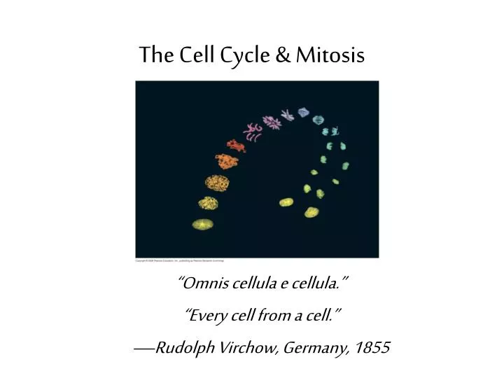
The Cell Cycle & Mitosis. “Omnis cellula e cellula.” “Every cell from a cell.” —Rudolph Virchow, Germany, 1855. Fig. 12-2a. Reproduction: An amoeba, A single celled eukaryote, divides into two cells. Each new cell will be an individual organism. 100 µm. Fig. 12-2b.

E N D
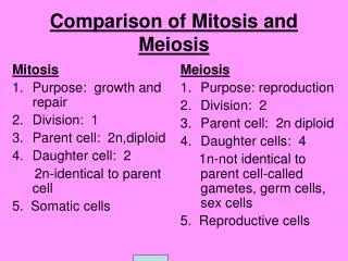
The Cell Cycle & Mitosis “Omnis cellula e cellula.” “Every cell from a cell.” —Rudolph Virchow, Germany, 1855
Fig. 12-2a Reproduction: An amoeba, A single celled eukaryote, divides into two cells. Each new cell will be an individual organism. 100 µm
Fig. 12-2b Growth and Development: A sand dollar embryo just after the fertilized egg divided. 200 µm
Fig. 12-2c Tissue Renewal: Bone marrow cells form new blood cells. 20 µm
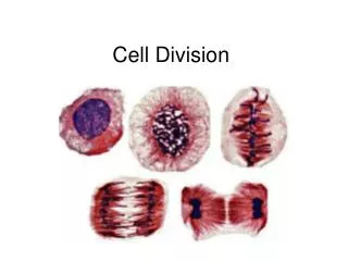
The Cell Cycle • The life of a cell from the time it first formed from a dividing parent cell until its own division into two cells. • Consists of interphase, mitosis & cytokinesis.
Interphase • The cell grows (increases in mass), • Copies cytoplasmic organelles and • Produces proteins • Duplicates chromosomes • 90% of cell cycle is in interphase
Mitosis Interphase Prophase Metaphase Anaphase Telophase Replication Alignment Separation
Fig. 12-6 Metaphase Anaphase Telophase and Cytokinesis G2 of Interphase Prophase Prometaphase Centrosomes (with centriole pairs) Early mitotic spindle Centromere Chromatin (duplicated) Fragments of nuclear envelope Nonkinetochore microtubules Aster Cleavage furrow Metaphase plate Nucleolus forming Daughter chromosomes Nuclear envelope forming Centrosome at one spindle pole Spindle Nuclear envelope Kinetochore Chromosome, consisting of two sister chromatids Kinetochore microtubule Plasma membrane Nucleolus
Replication • DNA replicates during interphase.
The Mitotic Spindle • Spindle fibers made of microtubules and proteins that controls chromosome movement during mitosis. • Assembled at centrosome in animal cells (MTOC)
Alignment (Prophase & Metaphase) • Microtubules extend from centrosomes and some attach to kinetochores on the chromatids • Microtublesmove the chromatids until their centromeres lie on the metaphase plate.
Separation—Anaphase & Telophase • Kinetochoremicrotubules shorten and pull the chromatids apart • The chromosomes are pulled to opposite poles of the cell
In animal cells, cytokinesis occurs by forming a cleavage furrow which deepens until the parent cell is pinched in two.
In plant cells, a cell plate forms in the middle of the cell.