Download
1 / 29
340 likes | 652 Views
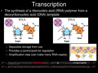
Activating Transcription. Chapter 25. 25.1 Introduction. Figure 25.1. 25.2 There Are Several Types of Transcription Factors. The basal apparatus determines the startpoint for transcription. Activators determine the frequency of transcription. Figure 25.2.

E N D
Activating Transcription Chapter 25
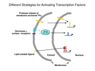
25.1 Introduction Figure 25.1
25.2 There Are Several Types of Transcription Factors • The basal apparatus determines the startpoint for transcription. • Activators determine the frequency of transcription. Figure 25.2
Activators work by making protein–protein contacts with the basal factors. • Activators may work via coactivators. • Some components of the transcriptional apparatus work by changing chromatin structure.
25.3 Independent Domains Bind DNA and Activate Transcription • DNA-binding activity and transcription-activation are carried by independent domains of an activator. • The role of the DNA-binding domain is to bring the transcription-activation domain into the vicinity of the promoter. Figure 25.3
25.4 The Two Hybrid Assay Detects Protein–Protein Interactions • The two hybrid assay works by requiring an interaction between two proteins: • one has a DNA-binding domain • the other has a transcription-activation domain Figure 25.6
25.5 Activators Interact with the Basal Apparatus • The principle that governs the function of all activators is: • A DNA-binding domain determines specificity for the target promoter or enhancer. • The DNA-binding domain is responsible for localizing a transcription-activating domain in the proximity of the basal apparatus. • An activator that works directly has: • a DNA-binding domain • an activating domain
An activator that does not have an activating domain may work by binding a coactivator that has an activating domain. Figure 25.7
Several factors in the basal apparatus are targets with which activators or coactivators interact. Figure 25.8
RNA polymerase may be associated with various alternative sets of transcription factors in the form of a holoenzyme complex. Figure 25.9
25.6 Some Promoter-Binding Proteins Are Repressors • Repression is usually achieved by affecting chromatin structure. • There are repressors that act by binding to specific promoters. Figure 25.10
25.7 Response Elements Are Recognized by Activators • Response elements may be located in promoters or enhancers. Figure 25.11
Each response element is recognized by a specific activator. • A promoter may have many response elements. • Elements may in turn activate transcription independently or in certain combinations.
25.8 There Are Many Types of DNA-Binding Domains • Activators are classified according to the type of DNA-binding domain. • Members of the same group have sequence variations of a specific motif that confer specificity for individual target sites.
25.9 A Zinc Finger Motif Is a DNA-Binding Domain • A zinc finger is a loop of ∼23 amino acids. • It protrudes from a zinc-binding site formed by His and Cys amino acids. • A zinc finger protein usually has multiple zinc fingers. Figure 25.13
The C-terminal part of each finger forms an α-helix. • The helix binds one turn of the major groove of DNA. • Some zinc finger proteins bind RNA instead of, or as well as, DNA. Figure 25.14
25.10 Steroid Receptors Are Activators • Steroid receptors are examples of ligand-responsive activators. • They are activated by binding a steroid (or other related molecules). • There are separate DNA-binding and ligand-binding domains. Figure 25.16
25.11 Steroid Receptors Have Zinc Fingers • The DNA binding domain of a steroid receptor is a type of zinc finger that has Cys but not His residues. • Glucocorticoid and estrogen receptors each have two zinc fingers. • The first determines the DNA target sequence. • Steroid receptors bind to DNA as dimers. Figure 25.17
25.12 Binding to the Response ElementIs Activated by Ligand-Binding • Binding of ligand to the C-terminal domain increases the affinity of the DNA-binding domain for its specific target site in DNA. Figure 25.19
25.13 Steroid Receptors Recognize ResponseElements by a Combinatorial Code • A steroid response element consists of two short half sites that may be palindromic or directly repeated. • There are only two types of half sites. • A receptor recognizes its response element by the orientation and spacing of the half sites.
The sequence of the half site is recognized by the first zinc finger. • The second zinc finger is responsible for dimerization, which determines the distance between the subunits. • Subunit separation in the receptor determines the recognition of spacing in the response element.
Some steroid receptors function as homodimers, whereas others form heterodimers. • Homodimers recognize palindromic response elements. • Heterodimers recognize response elements with directly repeated half sites. Figure 25.20 Figure 25.21
25.14 Homeodomains Bind Related Targets in DNA • The homeodomain is a DNA-binding domain of 60 amino acids that has three α-helices. Figure 25.24
The C-terminal α-helix-3 is 17 amino acids and binds in the major groove of DNA. • The N-terminal arm of the homeodomain projects into the minor groove of DNA. • Proteins containing homeodomains may be either activators or repressors of transcription. Figure 25.25
25.15 Helix-Loop-Helix Proteins Interact byCombinatorial Association • Helix-loop-helix proteins have a motif of 40 to 50 amino acids. • The motif comprises two amphipathic α-helices of 15 to 16 residues separated by a loop. • The helices are responsible for dimer formation. Figure 25.26
bHLH proteins have a basic sequence adjacent to the HLH motif that is responsible for binding to DNA. • Class A bHLH proteins are ubiquitously expressed. • Class B bHLH proteins are tissue-specific. • A class B protein usually forms a heterodimer with a class A protein.
HLH proteins that lack the basic region prevent a bHLH partner in a heterodimer from binding to DNA. • HLH proteins form combinatorial associations. • They may be changed during development by the addition or removal of specific proteins. Figure 25.27
25.16 Leucine Zippers Are Involved in Dimer Formation • The leucine zipper is an amphipathic helix that dimerizes. • The zipper is adjacent to a basic region that binds DNA.
Dimerization forms the bZIP motif: • The two basic regions symmetrically bind inverted repeats in DNA. Figure 25.28