Download
1 / 32
350 likes | 1.48k Views
Pacemaker for beginners. KITA yosuke Iizuka Hospital. Objectives. Review basic pacemaker terminology and function Discuss diagnosis and management of pacemaker emergencies . Historical Perspective. Electrical cardiac pacing for the management of brady-arrhythmias was first described in 1952

E N D
Pacemaker for beginners KITA yosuke Iizuka Hospital
Objectives • Review basic pacemaker terminology and function • Discuss diagnosis and management of pacemaker emergencies
Historical Perspective • Electrical cardiac pacing for the management of brady-arrhythmias was first described in 1952 • Permanent transvenous pacing devices were first introduced in the early 1960’s
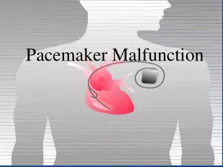
Pacemaker Components • Pulse Generator • Electronic Circuitry • Lead system
Pulse Generator • Lithium-iodine cell is the current standard battery • Advantages: • Long life – 4 to 10 years • Output voltage decreases gradually with time making sudden battery failure unlikely
Electronic Circuitry • Determines the function of the pacemaker itself • Utilizes a standard nomenclature for describing pacemakers
Lead Systems • Endocardial leads which are inserted using a subclavian vein approach • Actively fixed to the endocardium using screws or tines • Unipolar or bipolar leads
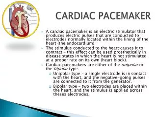
Electrocardiogram During Cardiac Pacing • Pacemaker has two main functions: • Sense intrinsic cardiac electrical activity • Electrically stimulate the heart • VVI- senses intrinsic cardiac activity in the ventricle and when a preset interval of time with no ventricular activity occurs it depolarizes the right ventricle causing ventricular contraction
Electrocardiogram • Dual chamber pacer is more complicated because the pacer has the ability to both sense and pace either the atrium or the ventricle • Possible to have only atrial, only ventricular or both atrial and ventricular pacing • DDD pacer is a common example of this
Atrial Spike Ventricular Spike
Ventricular Pacing AV Pacing
Magnet Placement • The EKG technician should perform a 12 lead cardiogram and then a rhythm strip with a magnet over the pacer • Often a very poorly understood concept by the non-cardiologist • Does not inactivate the pacer as is commonly believed • Activate a lead switch present in the pacemaker which converts the pacer to a asynchronous or fixed-rate pacing mode • Inhibits the sensing function of a pacemaker
Class I Indications For Permanent Pacing • Third degree AV block associated with: • Symptomatic bradycardia • Symptomatic bradycardia secondary to drugs required for dysrhythmia management • Asystole > 3 seconds or escape rate < 40 • After catheter ablation of the AV node • Post-op AV block not expected to resolve • Neuromuscular disease with AV block
Indications • Symptomatic bradycardia from second degree AV block • Bifascicular or trifascicular block with intermittent third degree or type II second degree block • Sinus node dysfunction with symptomatic bradycardia • Recurrent syncope caused by carotid sinus stimulation
Indications • Post myocardial infarction with any of: • Persistent second degree AV block with bilateral bundle branch block or third degree AV block • Transient second or third degree AV block and bundle branch block • Symptomatic, persistent second or third degree AV block
Infections • Pacemaker insertion is a surgical procedure: • 1% risk for bacteremia • 2% risk for wound or pocket infection • Usually occur soon after pacer insertion • Presence of a foreign body complicates management
Infection • Cellulitis or pocket infection: • Tenderness and redness over the pacemaker itself • Avoid performing a needle aspiration – damage the pacer • Bacteremia: Staphylococcus • aureus and Staphylococcus epi 60-70% of the time • Empiric antibiotics should include vancomycin pending culture
Infection • Consult the pacemaker physician • Draw blood cultures • Give appropriate antibiotics • Frequently the pacer and lead system need to be removed
Case 1 • 67 year old male presents to the emergency room 12 hours after insertion of a pacemaker complaining of left sided chest pain and shortness of breath • PR96, RR 33, BP 125/85, Oxygen saturation 88% RA • CXR as shown
Pneumothorax • Occurs during cannulation of the subclavian vien • Incidence - ?? Cardiologist dependent • Treatment: • Asymptomatic or small – observation • Symptomatic or large – tube thoracostomy • Notify the pacemaker physician
Case 2 • 72 year old male presents to the emergency room after a fall, tripped over a bath mat, no LOC • Shortened and rotated left leg • Past history – pacemaker, hypertension • Nurse does an routine pre-op CXR and EKG
Septal Perforation • Usually identified at the time of pacer insertion but leads can displace after insertion • Can occur with transvenous pacer insertion • Keys diagnosis are a RBBB pattern on EKG and a pacer lead displaced to the apex of the heart on CXR
Septal Perforation • Management: • Notify the pacer service • Pacer wire has to be removed but not emergently • Small VSD which heals spontaneously
Conclusions • Pacemakers are becoming more common everyday • We need to understand basic pacing terminology and modes to treat patients effectively. • Most pacer malfunctions are due to failure to sense, failure to capture, over-sensing, or in-appropriate rate • Standard ACLS protocols apply to all unstable patients with pacemakers.