Download
1 / 13

E N D
Cell-cell interaction The cytokines
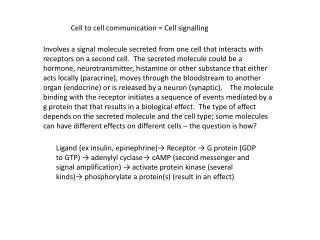
Definition • Cytokines are short-acting molecules that regulate the immune and inflammatory responses. Individual cytokines are often pleiotropic and have more than one function. Cytokines carry signals locally between cells, and thus have an effect on other cells.The term cytokine encompasses a large and diverse family of polypeptide regulators that are produced widely throughout the body by cells of diverse embryological origin. Each of the T-cell responses is dependent on cytokines produced by T-cells and other cells of the immune system. Cytokines are low molecular weight, secreted proteins that are produced transiently from appropriately stimulated cells.
Definition – con’d • Each cytokine has a matching cell-surface receptor. Subsequent cascades of intracellular signalling then alter cell functions. The effect of a particular cytokine on a given cell depends on the cytokine, its extracellular abundance, the presence and abundance of the complementary receptor on the cell surface, and downstream signals activated by receptor binding; these last two factors can vary by cell type. Cytokines are characterized by considerable "redundancy", in that many cytokines appear to share similar functions.
Cytokines of immunological significance • Interleukins (ILs) • Interferons (INFs) • Tumor Necrosis Factors (TNFs) • Chemokines
Interleukins (ILs) • A subset of cytokines numbered IL-l, IL-2, etc. • Particularly important in regulating the interactions between cells of the immune system. • They can bind to interleukin receptors (e.g. IL1RB2), chemokine receptors (CXCR1), or cluster of differentiation (e.g. CD212). • The vast majority of these are produced by T-helper cells. Other ILs are produced by monocytes, macrophages, and endothelial cells. They bind to receptors on their target cells and may stimulate the following activities: 1. Cell division, e.g. IL-2 on T-cells 2. Differentiation, e.g. IL-6 involved in differentiation of B-cells to plasma cells. 3. Activation of function, e.g. IL-4 is one of the second signals provided by the helper T-cell to the B-cell.
Interleukins - functions • IL-1: Th cell stimulation; B-cell maturation & proliferation; NK activation. • IL-2: produced exclusively by Th1 cells. Stimulates growth and differentiation of T-cells. • IL-3: stimulates differentiation and proliferation of myeloid progenitor cells. • IL-4: stimulates differentiation and proliferation of B-cells and proliferation of T-cells. • IL-6: stimulates plasmablasts to differentiate into plasma cells; initiates antibody secretion by plasma cells; stimulates the differentiation of haematopoietic stem cell; stimulates the differentiation of T-cells • IL-7: stimulates the differentiation and proliferation of lymphoid progenitor cells. • IL-8: stimulates Neutrophil chemotaxis. • IL-10: induces MФ to produce cytokines; B-cell activation; Th1 inhibition; Th2 activation. • IL-12: stimulates the differentiation of activated T-cells into Tc cells.
Interferons (IFNs) • A subset of cytokines named IFNα, IFNβ, IFNγ, etc • INF receptors are named IFNAR, IFNBR, IFNGR, etc • Although they are named after their ability to "interfere" with viral replication within host cells, IFNs have other functions: • They activate immune cells, such as natural killer cells and macrophages • They increase recognition of infection or tumor cells by up-regulating antigen presentation to T lymphocytes • They increase the ability of uninfected host cells to resist new infection by virus
Chemokines • Their name is derived from their ability to induce directed chemotaxis in nearby responsive cells; they are chemotactic cytokines. • Released by infected or damaged cells form a concentration gradient. Attracted cells move through the gradient towards the higher concentration of chemokine. • The major role of chemokines is to act as a chemo-attractant to guide the migration of cells. • Letters R and L may be added at the end of the nomenclature to differentiate whether it is a receptor or a ligand (e.g. CCL13, CCR13).
Chemokines - types • Members of the chemokine family are divided into four groups depending on the spacing of their first two cysteine residues (at the N terminal) into: • CC chemokines; proteins have 2 adjacent Cysteines • CXC chemokines; Cysteines are separated by one amino acid • C chemokines; only one Cysteine at N terminal • CX3C chemokines; have three amino acids between the two Cysteines
Tumor necrosis factors (TNFs) • A group of cytokines family that can cause cell death • A group of cytokines family that can cause cell death. TNFs act via the TNF Receptor (TNF-R) as a part of the extrinsic pathway for triggering apoptosis. TNF-R is associated with procaspases that can cleave other inactive procaspases and trigger the caspase cascade, irreversibly committing the cell to apoptosis. TNF interacts with tumor cells to trigger cytolysis or cell death • Are classified into TNFα and TNFβ
Cluster Of Differentiation (CD) • The cluster of differentiation is a protocol used for the identification and investigation of cell surface molecules present on white blood cells. CD molecules can act in numerous ways, often acting as receptors or ligands (the molecule that activates a receptor) important to the cell. A signal cascade is usually initiated, altering the behaviour of the cell. Some CD proteins do not play a role in cell signalling, but have other functions, such as cell adhesion. CD for humans is numbered up to 350 most recently. • The CD system is commonly used as cell markers, allowing cells to be defined based on what molecules are present on their surface. While using one CD molecule to define populations is uncommon, combining markers has allowed for cell types with very specific definitions.
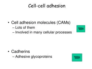
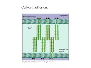
Cell adhesion molecules (CAMs) • Cell Adhesion Molecules (CAMs) are proteins located on the cell surface involved with the binding with other cells or with the extracellular matrix (ECM) in the process called cell adhesion. • Most of the CAMs belong to 5 protein families: immunoglobulin superfamily, integrins, cadherins, selectins and lymphocyte homing receptors. • The lymphocyte homing receptors are also called addressins. Two well known examples are CD34 and GLYCAM-1. • The three family members of selectins are E-selectin (endothelial), L-selectin (leukocyte), and P-selectin (platelet).