Download
1 / 81
820 likes | 1.28k Views
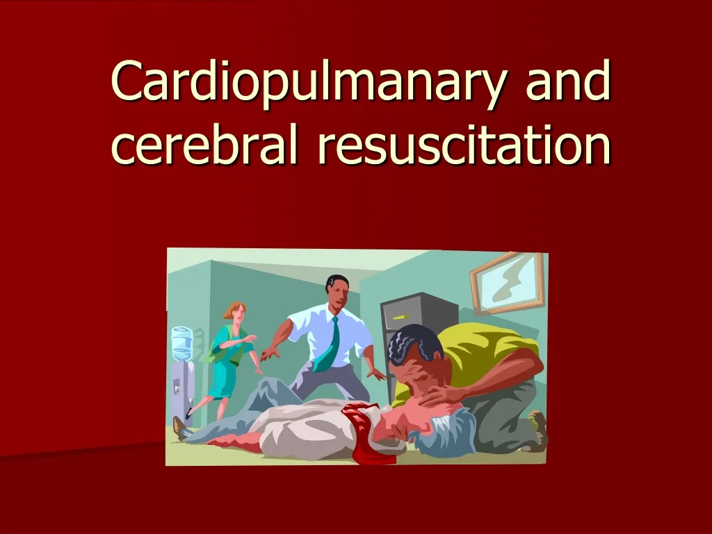
Cardiopulmanary and cerebral resuscitation. BLS. ALS. Basic Life Support Advanced Life Support Prolonged Life Support. AED. Survival Rate in Cardiopulmonary Arrest. 0 – 50 % (64%, 80%?). AED. Safety use Permissive legislation to use A ED (“Good Samaritan Law”)

E N D
BLS ALS Basic Life Support Advanced Life Support Prolonged Life Support AED
Survival Rate in Cardiopulmonary Arrest 0 – 50% (64%, 80%?)
AED Safety use Permissive legislation to use AED(“Good Samaritan Law”) 1 DEA – 10.000 inhabitants (population crowds,airports, police cars, fire fighters, casinos, etc.) An AED uses voice prompts to guide the rescuer. It analyses the ECG rhythm and informs the rescuer if a shock is needed. AEDs are extremely accurate and will deliver a shock only when VF (or its precursor, rapid ventricular tachycardia) is present .
Early BLS Done by lays (from bystanders) Until an emergency staff is available Double the surviving
DEFIBRILARE PRECOCE Fiecare 1 minut scade şansa de supravieţuire cu 7 - 10 % După 4 - 6 minute - leziuni neurologice După 10 minute - tentative de resuscitare nereuşite DEA ÎN 8-10 MINUTE!
SUPORTUL VITAL DE BAZASVB Scopul SVB: cardiac and cerebral oxygenation Increase the defibrillation efficiency DEFINITION: Basic life support (BLS) refers to maintaining airway patency and supporting breathing and the circulation, without the use of equipment other than a protective device
Consequences: - 3-5 min. => irreversible nerve cells lesions. Optmal time (until) to start CPR = 4 min. Longer time: - hypothermia (temp rectală 19-24 grade C) - barbiturics influence - child < 1 year old Shorter time, 2-3 min.: - respiratory arrest before heart stops ٠ barbiturics overdose ٠strangle
all cases accompanied with hypoxia extracardiac Causes of cardiac arrest cardiac Primary lesion of cardiac muscle leading to the progressive decline of contractility, conductivity disorders, mechanical factors
Cardiac ischemic heart disease (myocardial infarction, stenocardia) arrhythmias of different origin and character valvular disease cardiac tamponade pulmonary artery thromboembolism ruptured aneurysm of aorta reflector cardiac arrest Extracardiac airway obstruction acute respiratory failure shock electrolytic disorders embolisms of different origin drug overdose electrocution poisoning metabolic acidosis Causes of circulation arrest
FIOZIOPATOLOGIE Încetarea (ceasing) activit de pompă a cordului - lipsa debitului cardiac P.pf. miocardică şi coronariană => hipoxia cel, metabolism anaerob, acumularea de toxice lez tisulare ireversibile • vasodilataţie sitemică • vasoconstricţie pulm • rasp la catecolamine Acidoza P.pf. cerebrală = PAM – PIC (P sist) P.pf. miocard = PAM – P miocard (P diast)
DIAGNOSTIC of SCR • Rapid • unconscious + no respiration • + no pulse • ECG: asistole, FV, TV (activit electrică fără puls), PEA • Late clinical picture: - cyanosis and pale teguments • - areactive midriasis
BLS - sequence of operations • Check responsiveness • Call for help • Correctly place the victim and ensure the open airway • Check the presence of spontaneous respiration • Check pulse • Start external cardiac massage and artificial ventilation
In case of unconsciousness it is necessary to estimate quickly • the open airway • respiration • hemodynamics
Main stages of resuscitation A (Airway) – ensure open airway by preventing the falling back of tongue, tracheal intubation if possible B (Breathing) – start artificial ventilation of lungs C (Circulation) – restore the circulation by external cardiac massage D (Differentiation, Drugs, Defibrilation) – quickly perform differential diagnosis of cardiac arrest, use different medication and electric defibrillation in case of ventricular fibrillation
Probleme teoretice Ventilaţia artificială cu aer inspirat (16-18% O2) Teoria pompei cardiace - 30 % din perfuzia cerebrală optimă – limita critică a viabil. cel. corticale - fluxul miocardic – 20-30% din valoarea normală - fluxul visceral abdominal – 5%
1 Make sure you, the victim and any bystanders are safe. 2 Check the victim for a response Adult basic life support algorithm. • gently shake his shoulders and ask loudly: ‘‘Are you all right?’’ If he responds • leave him in the position in which you find him provided there is no further danger • try to find out what is wrong with him and get help if needed • reassess him regularly
If he does not respond • shout for help • turn the victim onto his back and then open the airway using head tilt and chin lift - place your hand on his forehead and gently tilt his head back keeping your thumb and index finger free to close his nose if rescue breathing is required. with your fingertips under the point of the victim’s chin, lift the chin to open the airway
Keeping the airway open, look, listen and feel for normal breathing • Look for chest movement. • Listen at the victim’s mouth for breath sounds. • Feel for air on your cheek. In the first few minutes after cardiac arrest, a victim may be barely breathing, or taking infrequent, noisy gasps. Do not confuse this with normal breathing. Look, listen, and feel for no more than 10 s to determine whether the victim is breathing normally. If you have any doubt whether breathing is normal, act as if it is not normal.
If he is breathing normally • turn him into the recovery position • send or go for help/call for an ambulance • check for continued breathing
If he is not breathing normally • send someone for help or, if you are on your own, leave the victim and alert the ambulance service; return and start chest compression as follows: . kneel by the side of the victim . place the heel of one hand in the centre of the victim’s chest. place the heel of your other hand on top of the first hand . interlock the fingers of your hands and ensure that pressure is not applied over the victim’s ribs. . position yourself vertically above the victim’s chest and, with your arms straight, .after each compression, release all the pressure on the chest without losing contact between your hands and the sternum; repeat at a rate of about 100/min. . . compression and release should take equal amounts of time
Combine chest compression with rescue breaths. • After 30 compressions open the airway again using head tilt and chin lift. • Pinch the soft part of the nose closed, using the index finger and thumb of your hand on the forehead. • Allow the mouth to open, but maintain chin lift. • Take a normal breath and place your lips around his mouth, making sure that you have a good seal. • Blow steadily into the mouth while watching for the chest to rise, taking about 1 s as in normal breathing; this is an effective rescue breath. • Maintaining head tilt and chin lift, take your mouth away from the victim and watch for the chest to fall as air passes out.
• Take another normal breath and blow into the victim’s mouth once more, to achieve a total of two effective rescue breaths. Then return your hands without delay to the correct position on the sternum and give a further 30 chest compressi. • Continue with chest compressions and rescue breaths in a ratio of 30:2. • Stop to recheck the victim only if he starts breathing normally; otherwise do not interrupt resuscitation. If your initial rescue breath does not make the chest rise as in normal breathing, then before your next attempt: • check the victim’s mouth and remove any obstruction • recheck that there is adequate head tilt and chin lift • do not attempt more than two breaths each time before returning to chest compr. If there is more than one rescuer present, another should take over CPR every 1—2 min to prevent fatigue. Ensure the minimum of delay during the changeover of rescuers
• Stop to recheck the victim only if he starts breathing normally; otherwise do not interrupt resuscitation. Continue resuscitation until • qualifed help arrives and takes over • the victim starts breathing normally • you become exhausted
Ventilation • During CPR the purpose of ventilation is to maintain adequate oxygenation. The optimal tidal volume, respiratory rate and inspired oxygen concentration • to achieve this, however, are not fully known. The current recommendations are based on the following evidence: • During CPR, blood flow to the lungs is substantially reduced, so an adequate ventilation perfusion ratio can be maintained with lower tidal volumes and respiratory rates than normal. • 2. Not only is hyperventilation (too many breaths or too large a volume) unnecessary, but it is harmful because it increases intrathoracic pressure, thus decreasing venous return to the heart and diminishing cardiac output. Survival is consequently reduced. • 3. When the airway is unprotected, a tidal volume of 1 l produces significantly more gastric distention than a tidal volume of 500 ml.
4. Low minute-ventilation (lower than normal tidal volume and respiratory rate) can maintain effective oxygenation and ventilation during CPR. During adult CPR, tidal volumes of approximately 500—600 ml (6—7 ml kg-1) should be adequate. 5. Interruptions in chest compression (for example to give rescue breaths) have a detrimental effect on survival. Giving rescue breaths over a shorter time will help to reduce the duration of essential interruptions. 6. The current recommendation is, therefore, for rescuers to give each rescue breath over about 1 s, with enough volume to make the victim’s chest rise, but to avoid rapid or forceful breaths. This recommendation applies to all forms of ventilation during CPR, including mouth-to-mouth and bagvalve-mask (BVM) with and without supplementary oxygen. 7. Mouth-to-nose ventilation is an effective alternative to mouth-to-mouth ventilation. It may be considered if the victim’s mouth is seriously injured or cannot be opened, the rescuer is assisting a victim in the water, or a mouth-to-mouth seal is difcult to achieve.
RESUSCITAREA EFECTUATĂ DE UN SINGUR SALVATOR RATA CTE : RESPIRAŢII 15:2 EFECTUATĂ DE DOI SALVATORI RATA CTE : RESPIRAŢII 5:1 RECOMANDAREA ANULUI 2002 RATA CTE : RESPIRAŢII 15:2 INDIFERENT DE NUMĂRUL DE SALVATORI RECOMANDAREA ANULUI 2006 RATA CTE : RESPIRAŢII 30:2 INDIFERENT DE NUMĂRUL DE SALVATORI
OBSTRUCŢIA CRS PRIN CORP STRĂIN RECUNOAŞTERE - TUSE - FLUX AERIAN BUN - PARŢIALĂ - EFORT RESPIRATOR • TUSE INEFICIENTĂ - FLUX AERIAN PROST • INSPIR ZGOMOTOS Noisy inspiration • CIANOZĂ - COMPLETĂ
EVALUAREA SEVERITĂŢII OBSTRUCŢIE SEVERĂ (tuse ineficientă) OBSTRUCŢIE UŞOARĂ (tuse eficientă) INCONŞTIENT CONŞTIENT RCP 5 LOVITURI INTERSCAPULARE 5 COMPRIMĂRI ABDOMINALE ÎNCURAJAREA TUSEI OBSTRUCŢIA CU CORP STRĂIN A CĂILOR AERIENE
ÎNDEPĂRTAREA OBSTRUCŢIEI 5 LOVITURI CU PODUL PALMEI LA NIVELUL SPAŢIULUI INTERSCAPULOVERTEBRAL 5 COMPRESIUNI ABDOMINALE MANEVRA HEIMLICH
MANEVRA HEIMLICH VICTIMĂ CONŞTIENTĂ VICTIMĂ INCONŞTIENTĂ
CPR Advanced Life Support
SUPORTUL VITAL AVANSAT • Scop:restore heart pomp activity by medication and defibrillation • - 8 min. since the heart stoped • maintaining BLS!! • IOT • venous acces • medication • DEFIBRILLATION !! – as soon as possible
FIBRILATIA VENTRICULARA = activitateaanarhica a maimultorcentriectopiciraspanditidifuz in micoardul ventricular; acesticentrigenereaza automatism producanddescarcarielectricezonaleceduc la contractiiparcelare, facandincapabilafunctia de pompa FV primara– aparepe un cord indemnhipoxic ( frecvent la copii) FV secundara– un mecanism de alteraremorfofunctionala a miocardului
TAHICARDIA VENTRICULARA = expresiaunordepolarizarisuccesive de origineventriculara (sub bifurcatiahisiana), de obiceicauzata de boalacornoranianaischemica TV nesustinuta< 30 sec. TV sustinuta> 30 sec. + colapshemodinamic In functie de morfologiacomplexului ORS clasificarea TV se va face in TV monomorfasi TV polimorfa O forma particulara de TV este torsada varfurilor generata de posdepolarizarea precoce in anumite conditii: QT lung (efect toxic a fenotiazidelor, antidepresive triciclice, haloperidol, antiaritmice), bradicardie severa, AVC-uri, dezechilibre hidroelectrolitice, hipotermie, boli cardiace
ASISTOLA • = lipsa totala a activitatii electrice a cordului, cu un prognostic rezervat, rata de supravietuire 1-2% • reprezentata de o linie sinusoidala (nu izoelectrica) compusa de mici unde date de depolarizarile de mica intensitate a musculaturii scheletice • trebuie diferentiata de FV cu unde mici
DISOCIATIA ELECTROMECANICA = entitatepatologicaparticulara a SCR, caracterizataprinasociereadintr-e o activitateelectricaprezenta (altadecat FV/TV) silipsaactivitatiimecanice a miocardului ventricular. DEM cu complexe QRS largi cu frecventascazutaapar in: IM masiv, hipopotasemiesevera, hipotermie, hipoxieacidoza, supradoajul de antidepresivetriciclice, beta-blocante, blocante ale canalelor de calciu, digitalice. DEM cu complexeingustecu frecventacrescuta (d.p.d.v. electric cordulraspunderelativ normal): hipovolemie, tampodanacardiaca, pneumotoracecompresiv, TEP masiv.
Shockable rhythms (ventricular fibrillation/pulseless ventricular tachycardia) * VF may be preceded by a period of VT or even supraventriculr tachycardia (SVT) VF/VT confirmed one shock (150 – 200 J biphasic) without reassessing the rhythm resume CPR (CV ration 30:2) 2 min. check monitor if there is still VF/VT second shock (200 J biphasic) resume CPR (CV ration 30:2) 2 min.
check monitor if there is still VF/VT Adrenaline 1 mg** 3rd shock (200 J biphasic) resumption of CPR for 2 min. analyze the rhythm if is still present VF/VT i.v. bolus of Amiodarone 300 mg 4th shock ** - adrenaline 1 mg every 3-5 min., once every two loops of the algorithm
Non-shockable rhythms (PEA and asystole) Asystole • PEA = pulseless electrical activity is defined as cardiac activity in the absence of any palpable pulses • mechanical myocardial contractions, but these are too weak to produce pulse • caused by reversible conditions CPR 30:2 Asystole/PEA i.v. + Adrenaline 1mg Atropine 3 mg secure airway, IOT Recheck the rhythm every 2 min
Airway and ventilation - without a good oxygenation it may be impossible to restore a spontaneous cardiac output. - consider reversible causes (4 H’s and 4 T’s) and, if identified, correct them. Tracheal intubation provides the most reliable healthcare provider but only for trained staff. - do not hyperventilate, ventilate the lungs at 10 breaths/min. Alternatives: - combitube - laryngeal mask airway (LMA) - laryngeal tube Deliver chest compressions, uninterrupted during ventilation.